Review
. 2013 Dec;7(6):449-54.
doi: 10.1007/s11832-013-0544-1. Epub 2013 Nov 29.

Longitudinal epiphyseal bracket

Affiliations
Review

Longitudinal epiphyseal bracket

Alexander D Choo et al. J Child Orthop. 2013 Dec.

Abstract

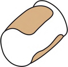
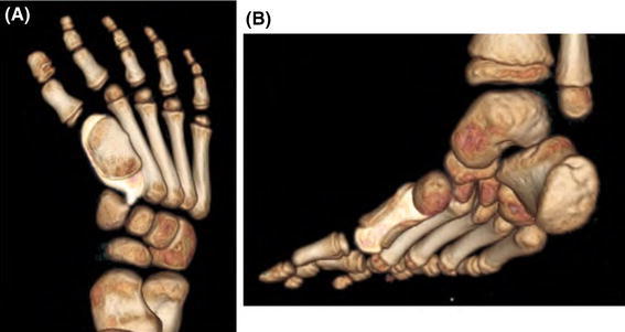
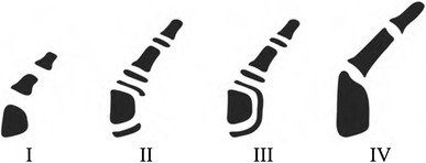
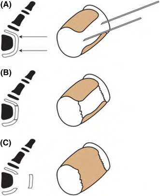
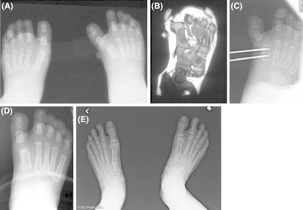
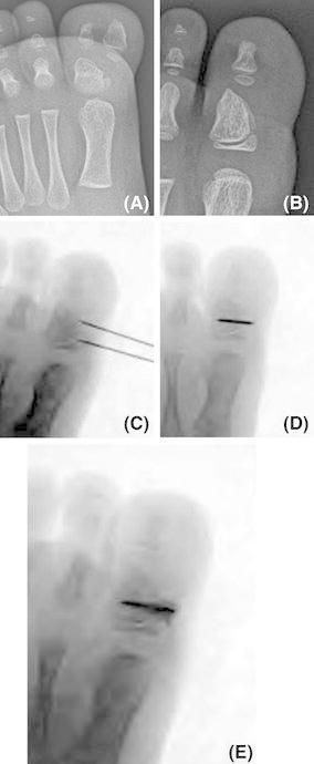
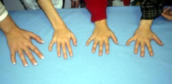
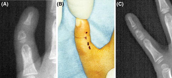
Longitudinal epiphyseal bracket or bracket epiphysis is an uncommon disorder of growth. Alternatively known as a delta phalanx, it is due to an anomalous secondary ossification center that extends longitudinally along the diaphysis. Although rare, longitudinal epiphyseal bracket most commonly manifests in the hands as clinodactyly and in the feet as hallux varus. Previously, longitudinal epiphyseal bracket has been treated with angular osteotomy, but we recommend early surgical physiolysis. We describe this uncommon disorder, our current recommendation for treatment, and present three illustrative cases.

Keywords: Bracket epiphysis; Clinodactyly; Delta phalanx; Hallux varus; Longitudinal epiphyseal bracket; Physiolysis.


Figures

Fig. 1
Fig. 1
Diagrammatic representation of longitudinal epiphyseal bracket. Note the abnormal secondary center of ossification along the diaphysis connecting the normal proximal and distal physis
Fig. 2
Fig. 2
Longitudinal epiphyseal bracket presenting as hallux varus. Three-dimensional reconstruction of foot CT of patient at age three with hallux varus demonstrates longitudinal epiphyseal bracket from both the (a) superior view and (b) posteromedial view of the foot
Fig. 3
Fig. 3
Radiographic classification of longitudinal epiphyseal bracket. Stage I is an ossified diaphysis with a radiolucent cartilaginous epiphysis. Stage II includes the appearance of secondary ossification centers at the epiphysis proximal and distal. In stage III there is ossification of the bracket epiphysis along the diaphysis. Stage IV, the final stage is closure of the bracket epiphysis
Fig. 4
Fig. 4
Diagrammatic representation for surgical physiolysis. a The border between the bracket and the proximal and distal epiphysis are marked with a small gauge needle and placement is checked on fluoroscopy. b The border of the bracket are sharply severed. c The bracket is excised, keeping normal epiphysis on the ends
Fig. 5
Fig. 5
Early presentation: unossified epipyseal bracket. a A 3-month-old male presents with a longitudinal epiphyseal bracket and unilateral hallux varus. Plain films demonstrate the D shaped metatarsal and polydactyl. b A MRI demonstrates the epiphyseal bracket. c At 4 months of age, he underwent excision of longitudinal epiphyseal bracket in the manner described. This intra-operative radiograph demonstrates the needles placed to mark the bracket resection area. Clinical and radiographic correction is noted at d age 2 years. At age 14 years his radiographs demonstrate his right first ray is nearly equal to his normal left side with normal alignment
Fig. 6
Fig. 6
Later presentation: ossified epiphyseal bracket. Illustration of an a ossified bracket with b initial treatment with bracket excision with Kirschner wire fixation and polymethylmethacrylate (PMMA) and c restoration of length and alignment with subsequent growth (From Mubarak et al. [10])
Fig. 7
Fig. 7
a A 2-year-old female with foot preaxial polydactyly. She underwent excision of bilateral preaxial polydactyly and reconstruction of the great toe at nineteen months. b At age two the patient is noted to have a bracket of the proximal phalanx with early ossification. The patient then underwent excision of the hallux bracket epiphysis. c Intra-operative fluoroscopy demonstrating placement of two needles at edges of bracket. d Following excision of the bracket, a Kirschner wire was placed which transversely through the proximal phalanx. e PMMA was then placed around the Kirschner wire
Fig. 8
Fig. 8
Clinical image of a family (parent and three children) demonstrating clinodactyly associated with longitudinal epiphyseal bracket of the hand little finger bilaterally
Fig. 9
Fig. 9
Surgical treatment of bracket of the finger in a 2-year-old female. a Radiographs of the left little finger note the short wedge middle phalanx. b Skin incision is marked along the radial aspect of the finger at the dorsal edge of the flexor crease from the proximal to distal interphalangeal joint. c Follow-up 3 years later shows improvement in length and less deformity

References

    1. Burnstein MI, De Smet AA, Breed AL, Thomas JR, Hafez GR. Longitudinal tibial epiphyseal bracket in Nievergelt syndrome. Skeletal Radiol. 1989;18:121–125. doi: 10.1007/BF00350660. - DOI - PubMed
    1. Elliott AM, Evans JA, Chudley AE, Reed MH. The duplicated longitudinal epiphysis or “kissing delta phalanx”: evolution and variation in three different disorders. Skeletal Radiol. 2004;33:345–351. doi: 10.1007/s00256-004-0752-3. - DOI - PubMed
    1. Jones GB. Delta phalanx. J Bone Joint Surg Br. 1964;46:226–228. - PubMed
    1. Jaeger M, Refior HJ. The congenital triangular deformity of the tubular bones of hand and foot. Clin Orthop Relat Res. 1971;81:139–150. doi: 10.1097/00003086-197111000-00023. - DOI - PubMed
    1. Carstam N, Theander G. Surgical treatment of clinodactyly caused by longitudinally bracketed diaphysis (“delta phalanx”) Scand J Plast Reconstr Surg. 1975;9:199–202. doi: 10.3109/02844317509022866. - DOI - PubMed

LinkOut - more resources