

Orthopaedic manifestations of chondroectodermal dysplasia: the Ellis-van Creveld syndrome
- PMID: 24432110
- PMCID: PMC3886354
- DOI: 10.1007/s11832-013-0541-4
Orthopaedic manifestations of chondroectodermal dysplasia: the Ellis-van Creveld syndrome
Abstract
Background: Ellis-van Creveld is a dwarfing syndrome transmitted as an autosomal recessive trait. The constant features of the condition include acromelic-micromelic dwarfism, ectodermal dysplasia involving the nails, teeth and gums, postaxial polydactyly of the hands and congenital heart disease. Congenital heart disease affects 50-60 % of all patients and nearly 50 % of patients die by 18 months of age from cardiopulmonary complications. This study is intended to characterise the orthopaedic manifestations of Ellis-van Creveld based on the authors' unique opportunity to interview and examine the largest group of patients to date in the literature.
Methods: Detailed interviews, physical examinations and/or radiographs were available on 71 cases of Ellis-van Creveld syndrome. Data were collected from physical examinations, radiographs, computed tomography (CT) reconstruction and magnetic resonance imaging (MRI) of the knee. Pathoanatomy of the knee was reinforced by the direct surgical observation of 25 limbs surgically managed during adolescence and puberty.
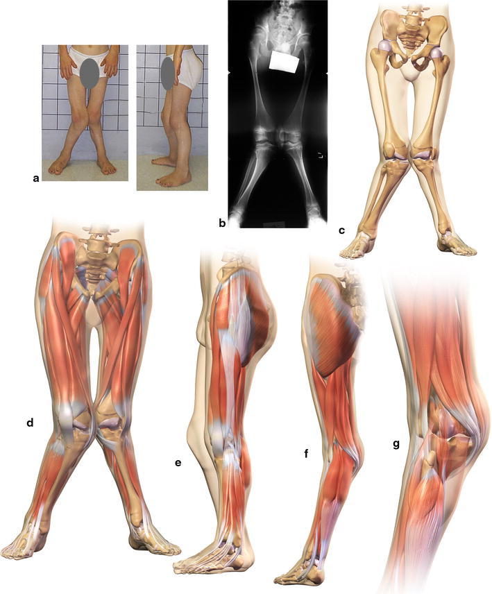
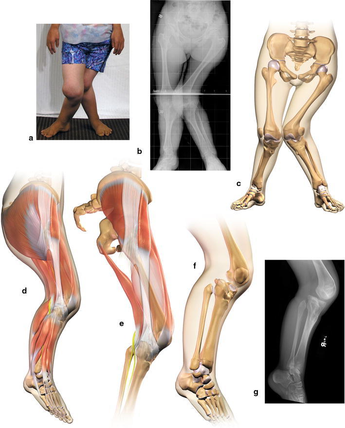
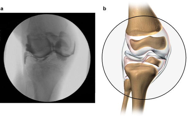
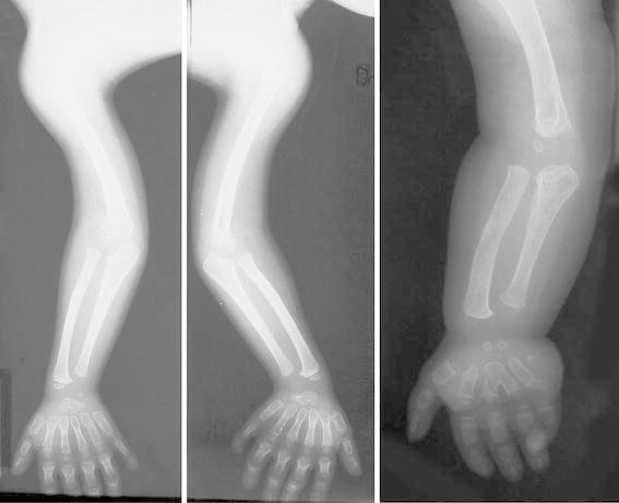
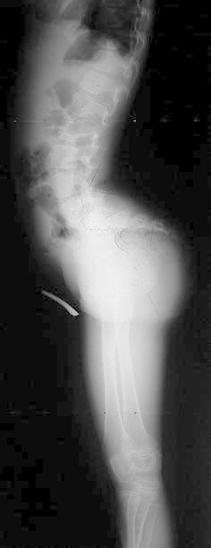
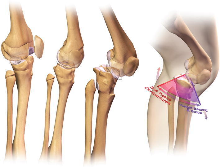
Results: A number of interesting clinical and radiographic abnormalities were noted in the upper extremities and lower extremities, but by far the most significant orthopaedic finding was a severe and relentlessly progressive valgus deformity of the knee. Although many patients had difficulties making a "fist" with the hand, no patient reported any functional disability. The severe valgus deformity of the knee is the result of a combination of profound contractures of the iliotibial band, lateral quadriceps, lateral hamstrings and lateral collateral ligament, leading to lateral patellar subluxation and dislocation. The lateral portion of the upper tibial plateau presents with cupping and progressive depression of the lateral plateau, along with severe valgus angulation of the proximal tibia and fibula. A proximal medial tibial exostosis is seen in nearly all cases.
Conclusion: This is the largest group of Ellis-van Creveld syndrome patients identified in the literature. An understanding of the orthopaedic pathoanatomy of the knee deformity is critical to determining the appropriate surgical management. This paper characterises the orthopaedic manifestations of Ellis-van Creveld syndrome and especially identifies the pathoanatomy of the severe and progressive valgus knee deformity.
Level of evidence: Level II.
Keywords: Chondroectodermal dysplasia; Ellis–van Creveld syndrome; Pathoanatomy.
Figures

Similar articles
-
An operative approach to address severe genu valgum deformity in the Ellis-van Creveld syndrome.J Child Orthop. 2014 Feb;8(1):61-9. doi: 10.1007/s11832-014-0552-9. Epub 2014 Jan 25. J Child Orthop. 2014. PMID: 24488845 Free PMC article.
-
Correction of knee deformity in patients with Ellis-van Creveld syndrome: A case report and review of the literature.Knee. 2012 Jun;19(3):218-22. doi: 10.1016/j.knee.2011.03.003. Epub 2011 Apr 5. Knee. 2012. PMID: 21470865
-
Ellis-van Creveld syndrome: a case report.J Pak Med Assoc. 2024 Feb;74(2):391-393. doi: 10.47391/JPMA.7049. J Pak Med Assoc. 2024. PMID: 38419244
-
Chondroectodermal dysplasia (Ellis van Creveld syndrome): a report of three cases with review of literature.Indian J Dent Res. 2007 Jan-Mar;18(1):31-4. doi: 10.4103/0970-9290.30920. Indian J Dent Res. 2007. PMID: 17347543 Review.
-
Rare clinical features of the Ellis van Creveld syndrome: A case report and literature review.Dermatol Ther. 2021 Jan;34(1):e14664. doi: 10.1111/dth.14664. Epub 2020 Dec 26. Dermatol Ther. 2021. PMID: 33314608 Review.
Cited by
-
Ellis-van Creveld Syndrome in Iran, a Case Report and Review of Disease Cases in Iran, Middle East.Acta Med Litu. 2021;28(2):317-324. doi: 10.15388/Amed.2021.28.2.11. Epub 2021 Aug 20. Acta Med Litu. 2021. PMID: 35474936 Free PMC article.
-
Successful Two-step Correction for Severe Genu Valgum in Ellis-van Creveld Syndrome: A Case Report.J Orthop Case Rep. 2017 Jul-Aug;7(4):13-16. doi: 10.13107/jocr.2250-0685.828. J Orthop Case Rep. 2017. PMID: 29181344 Free PMC article.
References
-
- McKusick VA, Hostetler JA, Egeland JA. Genetic studies of the Amish, background and potentialities. Bull Johns Hopkins Hosp. 1964;115:203–222. - PubMed
-
- McKusick VA, Egeland JA, Eldridge R, Krusen DE. Dwarfism in the Amish I. The Ellis–van Creveld syndrome. Bull Johns Hopkins Hosp. 1964;115:306–336. - PubMed
-
- McKusick VA, Egeland JA, Eldridge R, Krusen DE. Dwarfism in the Amish. I. The Ellis–van Creveld Syndrome. In: McKusick VA, editor. Medical genetic studies of the Amish: selected papers, assembled with commentary. Baltimore: John Hopkins University Press; 1978. pp. 93–117.
-
- McKusick VA, Hostetler J, Egeland JA, Eldridge R. Genetic studies of the Amish: background and potentialities. In: McKusick VA, editor. Medical genetic studies of the Amish: selected papers, assembled with commentary. Baltimore: John Hopkins University Press; 1978. pp. 1–22. - PubMed
-
- McKusick VA, Hostetler J, Egeland JA, Eldridge R. The distribution of certain genes in the Old Order Amish. In: McKusick VA, editor. Medical genetic studies of the Amish: selected papers, assembled with commentary. Baltimore: John Hopkins University Press; 1978. pp. 51–66.
LinkOut - more resources
Full Text Sources
Other Literature Sources
Research Materials
