

Reliability of assessing the coronal curvature of children with scoliosis by using ultrasound images
- PMID: 24432116
- PMCID: PMC3886351
- DOI: 10.1007/s11832-013-0539-y
Reliability of assessing the coronal curvature of children with scoliosis by using ultrasound images
Abstract
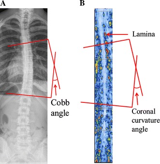
Purpose: To investigate the intra- and inter-observer reliability of the coronal curvature asymmetry of children with adolescent idiopathic scoliosis (AIS) using the center of lamina (COL) method on ultrasound (US) images.
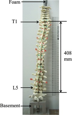
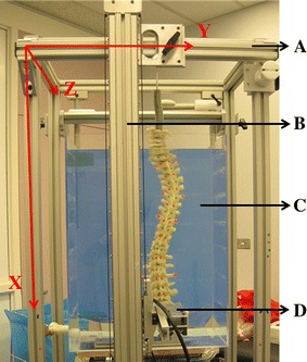
Methods: A cadaver spinal column phantom which was manipulated to present 30 scoliotic curves of varying severity of scoliotic deformities was scanned using both the US and laser scanner (LS) systems. Three observers of varying experience and skill measured the coronal curvature using the Cobb method on the LS images and the COL method on the US images. All of the measurements were performed twice, with a 1-week interval to reduce memory bias. The intra-class correlation coefficient (ICC), the mean absolute differences (MAD), and the error index (EI) were calculated to determine the agreement on selecting the end vertebrae. In addition, five AIS subjects were scanned using the US system. One observer measured the coronal curvature on the US images twice, and the measurements were compared with the Cobb angle reported in the clinical records.
Results: In the phantom study, the COL method showed high intra- and inter-observer reliabilities, with all ICC values >0.88. The maximum MAD of the COL measurements between different sessions among all observers was <4.1°. The EI values of the US method had similar end-vertebra selections as the LS method. The results of the pilot study showed a high intra-reliability for the US measurements. The measured difference between the Cobb and COL methods was 0.7° ± 0.5°.
Conclusions: The COL method using US images appears to be a very reliable method for measuring the coronal curvature in AIS without the need to expose the patient to radiation.
Keywords: Adolescent idiopathic scoliosis; Center of lamina method; Coronal curvature; Reliability; Ultrasound image.
Figures


Similar articles
-
Reliability and Validity Study of Clinical Ultrasound Imaging on Lateral Curvature of Adolescent Idiopathic Scoliosis.PLoS One. 2015 Aug 12;10(8):e0135264. doi: 10.1371/journal.pone.0135264. eCollection 2015. PLoS One. 2015. PMID: 26266802 Free PMC article.
-
Intra- and Inter-rater Reliability of Coronal Curvature Measurement for Adolescent Idiopathic Scoliosis Using Ultrasonic Imaging Method-A Pilot Study.Spine Deform. 2015 Mar;3(2):151-158. doi: 10.1016/j.jspd.2014.08.008. Epub 2015 Mar 4. Spine Deform. 2015. PMID: 27927306
-
3D ultrasound imaging provides reliable angle measurement with validity comparable to X-ray in patients with adolescent idiopathic scoliosis.J Orthop Translat. 2021 May 19;29:51-59. doi: 10.1016/j.jot.2021.04.007. eCollection 2021 Jul. J Orthop Translat. 2021. PMID: 34094858 Free PMC article.
-
Reliability of the axial vertebral rotation measurements of adolescent idiopathic scoliosis using the center of lamina method on ultrasound images: in vitro and in vivo study.Eur Spine J. 2016 Oct;25(10):3265-3273. doi: 10.1007/s00586-016-4492-6. Epub 2016 Mar 7. Eur Spine J. 2016. PMID: 26951170
-
Reliability and validity of lateral curvature assessments using clinical ultrasound for the patients with scoliosis: a systematic review.Eur Spine J. 2020 Apr;29(4):717-725. doi: 10.1007/s00586-019-06280-y. Epub 2020 Jan 10. Eur Spine J. 2020. PMID: 31925562
Cited by
-
Reliability and Validity Study of Clinical Ultrasound Imaging on Lateral Curvature of Adolescent Idiopathic Scoliosis.PLoS One. 2015 Aug 12;10(8):e0135264. doi: 10.1371/journal.pone.0135264. eCollection 2015. PLoS One. 2015. PMID: 26266802 Free PMC article.
-
Upright, prone, and supine spinal morphology and alignment in adolescent idiopathic scoliosis.Scoliosis Spinal Disord. 2017 Feb 22;12:6. doi: 10.1186/s13013-017-0111-5. eCollection 2017. Scoliosis Spinal Disord. 2017. PMID: 28251190 Free PMC article.
-
Reliability and accuracy of ultrasound measurements with and without the aid of previous radiographs in adolescent idiopathic scoliosis (AIS).Eur Spine J. 2015 Jul;24(7):1427-33. doi: 10.1007/s00586-015-3855-8. Epub 2015 Mar 10. Eur Spine J. 2015. PMID: 25753005
-
Comparison of ultrasound imaging and cone-beam computed tomography for examination of the alveolar bone level: A systematic review.PLoS One. 2018 Oct 3;13(10):e0200596. doi: 10.1371/journal.pone.0200596. eCollection 2018. PLoS One. 2018. PMID: 30281591 Free PMC article.
-
A reliability and validity study for Scolioscan: a radiation-free scoliosis assessment system using 3D ultrasound imaging.Scoliosis Spinal Disord. 2016 May 31;11:13. doi: 10.1186/s13013-016-0074-y. eCollection 2016. Scoliosis Spinal Disord. 2016. PMID: 27299162 Free PMC article.
References
-
- Cobb J (1948) Outline for the study of scoliosis. In: The American Academy of Orthopaedic Surgeons. Instructional Course Lectures, vol 5. Edwards, Ann Arbor, pp 261–275
-
- Helenius I, Remes V, Yrjonen T, Ylikoski M, Schlenzka D, Helenius M, Poussa M. Harrington and Cotrel-Dubousset instrumentation in adolescent idiopathic scoliosis. Long-term functional and radiographic outcomes. J Bone Joint Surg Am. 2003;85A(12):2303–2309. - PubMed
-
- Carman DL, Browne RH, Birch JG. Measurement of scoliosis and kyphosis radiographs. Intra-observer and inter-observer variation. J Bone Joint Surg Am. 1990;72(3):328–333. - PubMed
LinkOut - more resources
Full Text Sources
Other Literature Sources
Medical
