

Review
doi: 10.1007/s12154-013-0094-5.
Fluorescent labeling and modification of proteins
Affiliations
- PMID: 24432126
- PMCID: PMC3691395
- DOI: 10.1007/s12154-013-0094-5
Item in Clipboard
Review
Fluorescent labeling and modification of proteins
J Chem Biol.
.
Abstract
This review provides an outline for fluorescent labeling of proteins. Fluorescent assays are very diverse providing the most sensitive and robust methods for observing biological processes. Here, different types of labels and methods of attachment are discussed in combination with their fluorescent properties. The advantages and disadvantages of these different methods are highlighted, allowing the careful selection for different applications, ranging from ensemble spectroscopy assays through to single-molecule measurements.
Keywords: Fluorescence; Fluorescent proteins; Fluorophores; Quantum dots; Single molecule.
Figures

Methods for fluorescent labeling of proteins. a A generic scheme for labeling the protein of interest (POI). A fluorophore is attached to reactive group A. The complementary reactive group B is attached to the POI for labeling. b Reaction with a maleimide-conjugated fluorophore and cysteine residue on POI. c Reaction with a succinimidyl-ester conjugated fluorophore and N-terminal amine on POI. d Example reaction of peptide ligation. A thioester on the POI is ligated to a fluorescent peptide through an N-terminal cysteine residue. e Example of a self-labeling tetracysteine tag which binds to the bis-ARSenic fluorescein (FlAsH) probe. f Example of a self-labeling protein tag, the SNAP-Tag. The fluorescent O6-benzylguanine is cleaved by hAGT resulting in the fluorophore being covalently linked to the hAGT and POI. g Biotinylation of the POI at the biotin recognition sequence (BRS) by BirA and conjugation with streptavidin–fluorophore or functionalized quantum dot
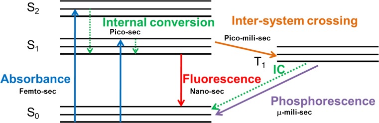
Excitation and emission of a fluorophore. Modified Jablonski diagram representing the excitation and emission cycle of a fluorophore
References
Publication types
LinkOut - more resources
Full Text Sources
Other Literature Sources
