

Renal artery aneurysm mimicking a solid parenchymal lesion
- PMID: 24432164
- PMCID: PMC3771572
- DOI: 10.1007/s40477-013-0021-1
Renal artery aneurysm mimicking a solid parenchymal lesion
Abstract
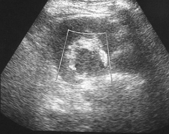
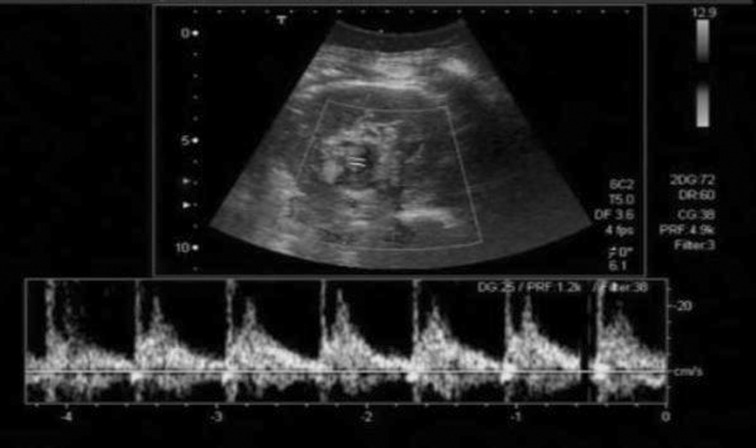
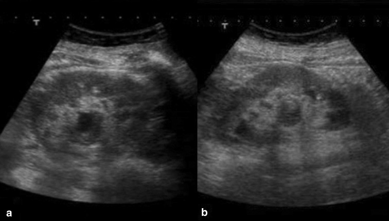
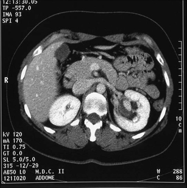
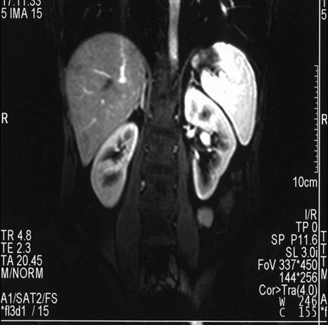
A 40-year-old woman was referred to our department for further investigation of a renal mass identified at an ultrasound (US) examination carried out in a private clinic because of abdominal pain. The mass was oval and hypoechoic, measured about 20 mm in diameter and was located near the left renal sinus; color Doppler showed peripheral blood flow. US examination carried out in our department using different equipment confirmed the presence of the mass but revealed intralesional blood flow suggesting aneurysm. This diagnosis was confirmed at subsequent computed tomography (CT) scanning and magnetic resonance imaging (MRI). The patient refused to undergo surgery and she is currently being monitored and has suffered no sequelae.
Una donna di 40 anni giungeva alla nostra osservazione per la rivalutazione di una formazione renale identificata ad un esame ecografico effettuato esternamente per dolore addominale. Tale formazione, ovalare ipoecogena di circa 20 mm, era localizzata a livello del seno renale di sinistra e mostrava al color Doppler segnale vascolare perifericamente. Il controllo ecografico da noi eseguito con apparecchiatura più performante confermava la presenza della formazione evidenziando tuttavia segnale vascolare al suo interno e ponendo il sospetto di aneurisma, confermato poi ai successivi esami TC ed RM. La paziente rifiutava di sottoporsi ad intervento chirurgico terapeutico ed è tuttora in follow-up senza sequele.
Keywords: Aneurysm; Color Doppler; Renal mass; Ultrasound.
Figures
Similar articles
-
Regarding six cases of mesenteric panniculitis: US, spiral CT, Magnetic Resonance.Radiol Med. 2002 May-Jun;103(5-6):511-8. Radiol Med. 2002. PMID: 12207186 English, Italian.
-
[Adrenal hemorrhage in a patient with systemic lupus erythematosus].Beijing Da Xue Xue Bao Yi Xue Ban. 2019 Dec 18;51(6):1178-1181. doi: 10.19723/j.issn.1671-167X.2019.06.036. Beijing Da Xue Xue Bao Yi Xue Ban. 2019. PMID: 31848526 Free PMC article. Chinese.
-
Multiple visceral artery aneurysms.Ann Vasc Surg. 2015 Aug;29(6):1318.e7-1318.e10. doi: 10.1016/j.avsg.2015.02.026. Epub 2015 Jun 12. Ann Vasc Surg. 2015. PMID: 26072724
-
[A case of retroperitoneal venous aneurysm].Hinyokika Kiyo. 1992 Sep;38(9):1037-40. Hinyokika Kiyo. 1992. PMID: 1414755 Review. Japanese.
-
[Integrated diagnosis of liver angioma: comparison of Doppler color ultrasonography, computerized tomography, and magnetic resonance].Radiol Med. 1997 Jan-Feb;93(1-2):87-94. Radiol Med. 1997. PMID: 9380876 Review. Italian.
Cited by
-
Giant Renal Artery Aneurysm With Hydronephrosis and Severe Atrophy of the Renal Parenchyma: Case Report and Literature Review.Clin Med Insights Case Rep. 2022 Oct 8;15:11795476221127129. doi: 10.1177/11795476221127129. eCollection 2022. Clin Med Insights Case Rep. 2022. PMID: 36225860 Free PMC article.
-
Large renal artery aneurysm masquerading as renal cell carcinoma.Urol Case Rep. 2022 May 21;43:102117. doi: 10.1016/j.eucr.2022.102117. eCollection 2022 Jul. Urol Case Rep. 2022. PMID: 35646597 Free PMC article.
References
-
- Shonai T, Koito K, Ichimura T, Hirokawa N, Sakata K, Hareyama M. Renal artery aneurysm: evaluation with color Doppler ultrasonography before and after percutaneous transarterial embolization. J Ultrasound Med. 2000;19:277–280. - PubMed
LinkOut - more resources
Full Text Sources
Other Literature Sources
