Review
. 2013 Nov 26;16(4):179-86.
doi: 10.1007/s40477-013-0049-2.

Acute nontraumatic liver lesions

Affiliations
Review

Acute nontraumatic liver lesions

Marcello Caremani et al. J Ultrasound. .

Abstract

The principal conditions requiring emergency/urgent intervention in patients with nontraumatic liver lesions are hemorrhage (with or without tumor rupture), rupture of hydatid cysts (with or without infection), complications arising from liver abscesses or congenital liver cysts, rupture related to peliosis hepatis, and in rare cases spontaneous hemorrhage. This article examines each of these conditions, its appearance on ultrasound (the first-line imaging method of choice for assessing any urgent nontraumatic liver lesion) and indications for additional imaging studies.

Le principali cause di emergenza/urgenza in patologia epatica non traumatica sono l’emorragia con o senza rottura di neoplasie epatiche, la rottura con o senza infezione delle cisti idatidee, le complicanze degli ascessi epatici, delle cisti congenite del fegato, l’emorragia, la rottura in corso di peliosi epatica e la rara emorragia spontanea. Dopo aver esaminato singolarmente le varie cause di emergenza/urgenza, i rispettivi aspetti ecografici e l’integrazione con altre metodiche di imaging si conclude che nei pazienti con patologia epatica l’ecografia, con o senza mezzo di contrasto, è la metodica da utilizzare in prima istanza ad ogni emergenza/urgenza non traumatica del fegato.

Keywords: Acute liver disorders; Contrast-enhanced ultrasound; Ultrasound.


Figures

Fig. 1
Fig. 1
Ruptured hepatocellular carcinoma. Large, inhomogeneous lesion located in the left lobe of the liver in a patient with cirrhosis (a). Administration of Sonovue (b) reveals avascular areas within the lesion characterized by little or no contrast medium
Fig. 2
Fig. 2
Contrast-enhanced ultrasound imaging of a focal liver lesion revealed a hepatic adenoma containing avascular areas caused by tumor rupture
Fig. 3
Fig. 3
Large angioma of the right lobe of the liver in a patient hospitalized for intense right hypochondriac pain (a). CEUS reveals large avascular areas caused by acute thrombosis (b)
Fig. 4
Fig. 4
Rupture of a giant angioma. The large inhomogeneous lesion that occupies the entire right lobe of the liver represents a giant angioma with a V-shaped anechoic area that extends all the way to the Glisson capsule (a). Perihepatic liquid represents blood in the peritoneum (b)
Fig. 5
Fig. 5
Spontaneous rupture of a liver abscess. Contrast-enhanced ultrasound of the liver in a patient with a liver abscess shows an avascular area representing the abscess, interruption of the liver capsule, and a small fluid collection around the Glisson capsule (arrows)
Fig. 6
Fig. 6
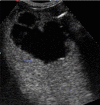
Large, nonparasitic liver cyst with fine, stratified echoes posteriorly representing spontaneous intracystic hemorrhage
Fig. 7
Fig. 7
Suppuration of a hydatid cyst caused by gas-producing microbes. Complex hepatic lesion with internal reverberation artifacts (a). b, d Small daughter cysts and the wall of the lesion itself can also be seen (c)
Fig. 8
Fig. 8
Rupture of a hydatid cyst in the bile ducts. The large hydatid cyst is in contact with the right bile duct (a). The main bile duct and the gallbladder both contain echogenic material without posterior shadowing (b)

Similar articles

Cited by

References

    1. Yeh CN, Lee WC, Jeng LB, Chen MF, Yu MC. Spontaneous tumor rupture and prognosis in patients with hepatocellular carcinoma. Br J Surg. 2002;89(9):1125–1129. doi: 10.1046/j.1365-2168.2002.02188.x. - DOI - PubMed
    1. Vergara V, Muratore A, Bouzari H, Polasti R, Ferrero A, Galatola G, Capussotti L. Spontaneous rupture of hepatocellular carcinoma: surgical resection. Eur J Surg Oncol. 2000;26(8):770–772. doi: 10.1053/ejso.2000.1001. - DOI - PubMed
    1. Vivarelli M, Cavallari A, Bellusci R, De Raffele E, Nardo B, Gozzetti G. Ruptured hepatocellular carcinoma:an important cause of haemoperitoneum. Eur Surg. 1995;161(2):881–886. - PubMed
    1. Chen CY, Lin XZ, Shin JS, Lin CY, Leow TC, Chen CY, Chang TT. Spontaneous rupture of hepatocellular carcinoma. J Clin Gastroenterol. 1995;21(3):238–242. doi: 10.1097/00004836-199510000-00015. - DOI - PubMed
    1. Zhu LX, Wang GS, Fan ST. Spontaneous rupture of hepatocellular carcinoma. Br J Surg. 1996;83(5):602–607. doi: 10.1002/bjs.1800830507. - DOI - PubMed

LinkOut - more resources