Review
. 2013 Mar 2;16(4):215-22.
doi: 10.1007/s40477-013-0010-4.

Complications of muscle injuries

Affiliations
Review

Complications of muscle injuries

F Alessandrino et al. J Ultrasound. .

Abstract

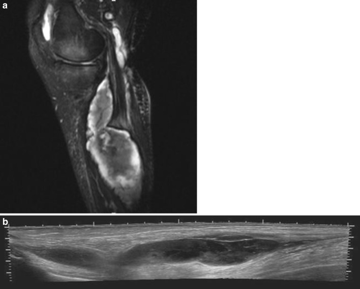
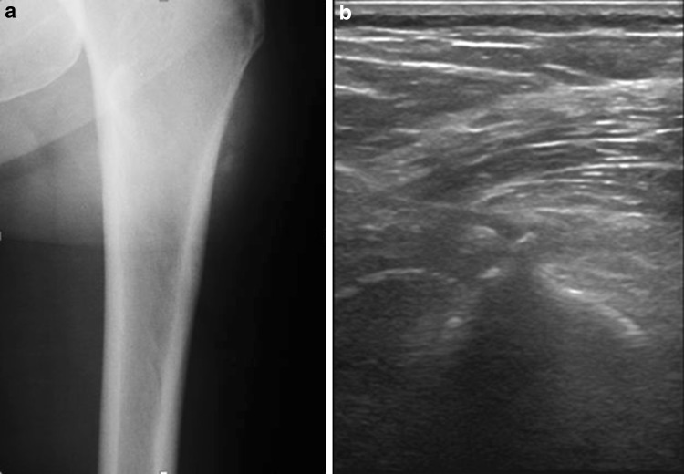
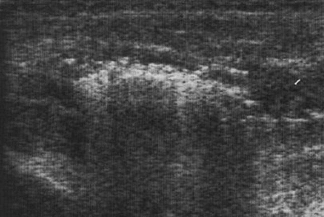
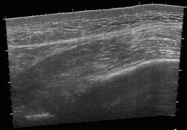
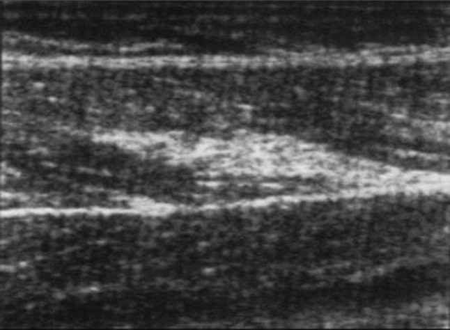
Muscle injuries can be classified into strain injuries and contusions. Depending on the type of injury, different complications may occur, which in turn can be divided into early, intermediate and delayed complications. A prompt diagnosis of complications allows early treatment and permits to avoid harmful sequelae. Imaging studies, ultrasonography in particular, allow (recognizing) the assessment of complications whenever clinically suspected. In this article the most frequent complications of muscle injuries are presented.

Le lesioni muscolari possono essere classificate in lesioni contusive e lesioni distrattive. A seconda del tipo di lesione, possono insorgere complicanze differenti, che a loro volta possono essere suddivise in complicanze precoci, intermedie e tardive. Un rapido e preciso riconoscimento delle complicanze consente di intervenire precocemente e permette di evitare conseguenze dannose. L’imaging, in particolar modo l’ecografia, permette il riconoscimento di complicanze ogni qualvolta esse siano sospettate clinicamente. In questo articolo vengono presentate le complicanze più frequenti delle lesioni muscolari.

Keywords: Complications; Muscle injuries; Ultrasound.


Figures

Fig. 1
Fig. 1
a Sagittal sonogram of the posterior lower leg demonstrating an acute gastrocnemic vein thrombosis. The thrombus is hypoechoic, filling the central portion of the vein. b Colour Doppler demonstrates venous flow only in the peripheral part of the vein, while the central part is occupied by the thrombus
Fig. 2
Fig. 2
a Sagittal fat saturated T2-weighted magnetic resonance image of the lower leg demonstrating a ruptured baker cyst which eventually caused an acute compartmental syndrome of the lower leg. b Extended field-of-view longitudinal sonogram of the same patient, where debris can be observed within the ruptured cyst
Fig. 3
Fig. 3
a Radiograph of a left femur showing oval-shaped extraskeletal calcifications. b Sagittal sonogram of the same patient showing homogenous, hyperechoic, well-defined oval-shaped masses, with acoustic shadowing
Fig. 4
Fig. 4
Transverse sonogram of a patient with pyomyositis of the rectus femoris muscle. Diffuse muscle swelling and partial posterior acoustic enhancement can be seen, suggesting a gas-forming organism infection
Fig. 5
Fig. 5
Sagittal extended field-of-view sonographic image of the right leg of a patient with rhabdomyolisis. Diffuse muscle swelling with loss of muscle striations can be seen
Fig. 6
Fig. 6
Sagittal sonogram of the left leg where a hyperechoic linear lesion attached to the epymisium of the rectus femoris can be seen consistent with posttraumatic fibrosis
Fig. 7
Fig. 7
a Patient with a lower leg muscle hernia. A lump can be observed in the lateral part of the lower leg. b Sagittal sonogram of the same patient. Muscle tissue can be seen protruding through an epymisial defect

References

    1. Järvinen TA, Järvinen TL, Kääriäinen M, Kalimo H, Järvinen M. Muscle injuries: biology and treatment. Am J Sports Med. 2005;33:745–764. doi: 10.1177/0363546505274714. - DOI - PubMed
    1. Orchard J, Best TM. The management of muscle strain injuries: an early return versus the risk of recurrence. Clin J Sport Med. 2002;12:3–5. doi: 10.1097/00042752-200201000-00004. - DOI - PubMed
    1. Orchard J, Best TM, Verrall GM. Return to play following muscle strains. Clin J Sport Med. 2005;15:436–441. doi: 10.1097/01.jsm.0000188206.54984.65. - DOI - PubMed
    1. Flecca D, Tomei A, Ravazzolo N, Martinelli M, Giovagnorio F. US evaluation and diagnosis of rupture of the medial head of the gastrocnemius (“Tennis Leg”) J Ultrasound. 2007;10(4):194–198. doi: 10.1016/j.jus.2007.09.007. - DOI - PMC - PubMed
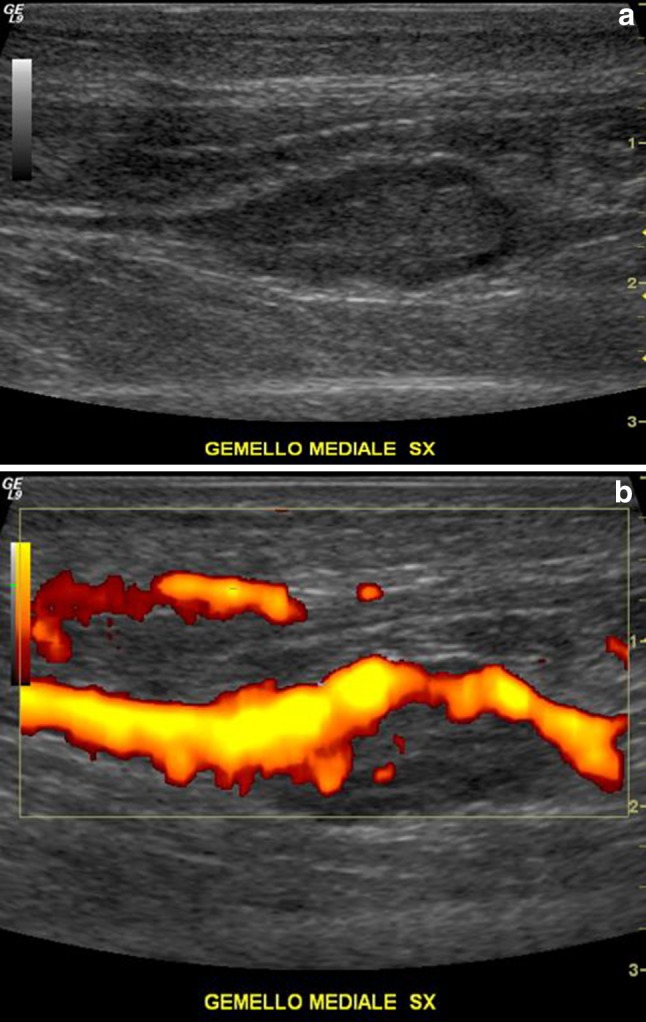
    1. Bianchi S, Martinoli C, Abdelwahab IF, Derchi LE, Damiani S. Sonographic evaluation of tears of the gastrocnemius medial head (“tennis leg”) J Ultrasound Med. 1998;17(3):157–162. - PubMed