

doi: 10.1016/j.mmcr.2013.02.008.
Mind the gap: Management of an emergent and threatening invasive fungal infection-a case report of rhino-orbital-cerebral and pulmonary mucormycosis
Affiliations
- PMID: 24432223
- PMCID: PMC3885946
- DOI: 10.1016/j.mmcr.2013.02.008
Item in Clipboard
Mind the gap: Management of an emergent and threatening invasive fungal infection-a case report of rhino-orbital-cerebral and pulmonary mucormycosis
Med Mycol Case Rep.
.
Abstract
Mucormycosis is an emergent and threatening invasive fungal invasion underdiagnosed by clinicians due to lack of awareness and aspecific clinical picture. The authors describe a clinical case of a diabetic and cirrhotic patient who developed rhino-orbital-cerebral and pulmonary mucormycosis, non-responsive to treatment. Typical gaps in the management of this deadly disease are addressed. There is a strong need for novel therapies and an expectation that sponsors will recognize the critical need for randomized clinical trials.
Keywords: Delays; Diabetes; High mortality; Hyperbaric medicine; Mucormycosis; Randomized clinical trials.
Figures
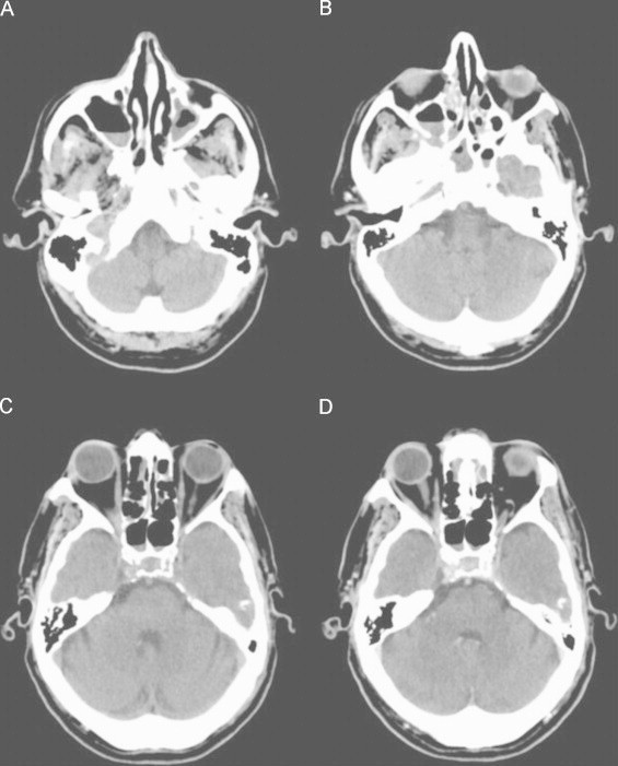
Cerebral CT scan, axial images. (A–C), Non-enhanced CT scan; (D) Contrast-enhanced CT scan. Partial soft tissue opacification of the maxillary, sphenoid and ethmoid sinuses.
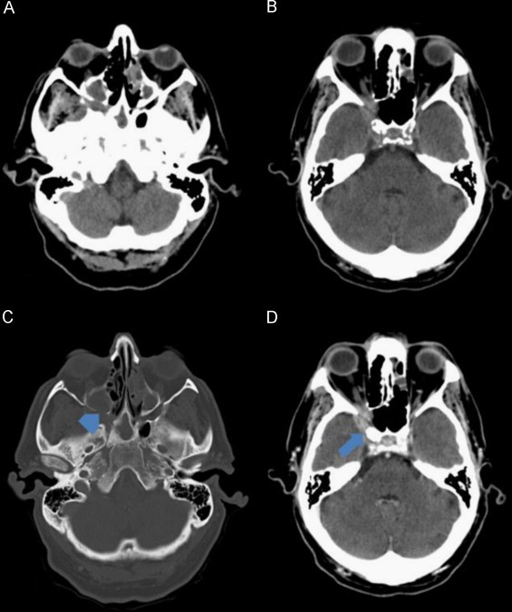
Cerebral CT scan, axial images. (A and B) Non-enhanced CT scan; (C) CT scan bone window; (D) Contrast-enhanced CT scan. Soft tissue opacification of the paranasal sinuses associated with focal areas of bone erosion of the posterior wall of the right maxillary sinus (arrowhead). Extension of the soft tissue lesion to the pterygopalatine fossa and intracranial extension to the temporal fossa (arrow) and to the anterior part of the cavernous sinus.
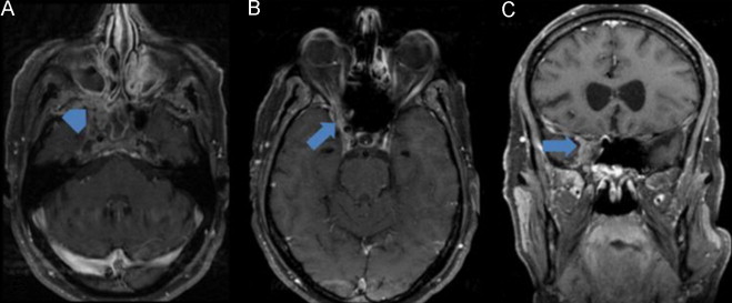
Cerebral MRI: (A, B) (axial T1 with fat saturation after gadolinium) and (C) (coronal T1 with fat saturation after gadolinium). Lesion centered on the apex of the right orbit, filling the sphenoidal fissure and optic canal, extending to the pterygopalatine fossa (arrowhead) and with mild intracranial extra-axial extension (arrow). Lesion is isointense in T1 and hypointense in T2 (not shown) with homogeneous enhancement after gadolinium. Associated inflammatory changes of the sinuses are also observed.
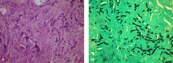
(A) Hematoxylin and eosin stain, 400× and (B) Grocott's methenamine silver stain, 400×. Multiple broad non-septated, ribbon-like, right-angle hyphae.
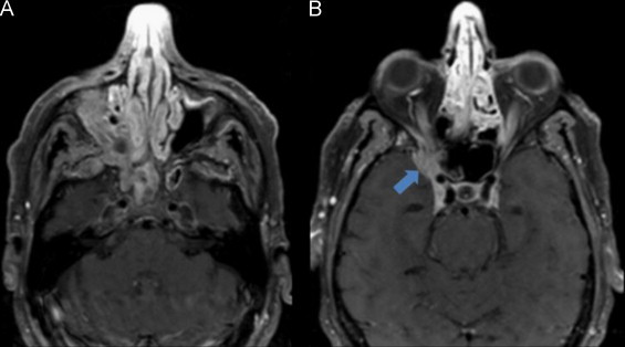
(A and B) (Axial T1 with fat saturation after gadolinium) Cerebral MRI at day 20 of Liposomal amphotericin B and caspofungin treatment, showing imagiological worsening with increased intracranial infiltration of the fungal lesion (arrow).
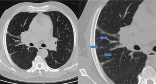
Thorax CT scan: muliple peripherical centrilobular nodules with “tree-in-bud” images (arrows), showing endobronchial spread of infection.
References
-
- Marques DS, Pinho Vaz C, Branca R, Campilho F, Lamelas C, Afonso LP et al. Rhizomucor and scedosporium infection post hematopoietic stem-cell transplant. Case Report Med 2011;830769. doi:10.1155/2011/830769. Epub 2011 April 5. - DOI - PMC - PubMed
-
- Oliveira V., Costa A. Cerebral hematoma caused by mucormycosis. Reviews in Neurological Diseases. 2001;33(10):951–953. - PubMed
-
- Rammaert B., Lanternier F., Poirée S., Kania R., Lortholary O. Diabetes and mucormycosis: a complex interplay. Diabetes and Metabolism. 2012;38(3):193–204. - PubMed
LinkOut - more resources
Full Text Sources
Other Literature Sources
Miscellaneous
