

Motor recovery and microstructural change in rubro-spinal tract in subcortical stroke
- PMID: 24432247
- PMCID: PMC3891492
- DOI: 10.1016/j.nicl.2013.12.003
Motor recovery and microstructural change in rubro-spinal tract in subcortical stroke
Abstract
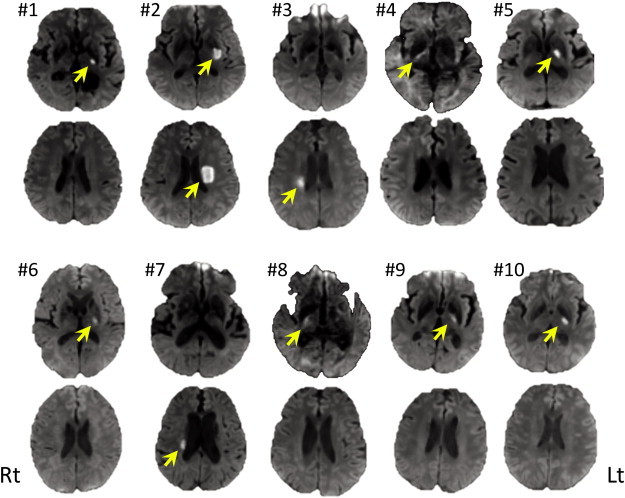
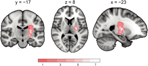
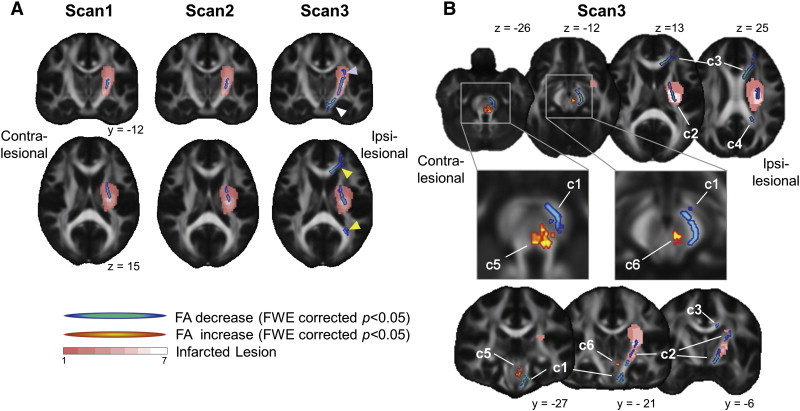
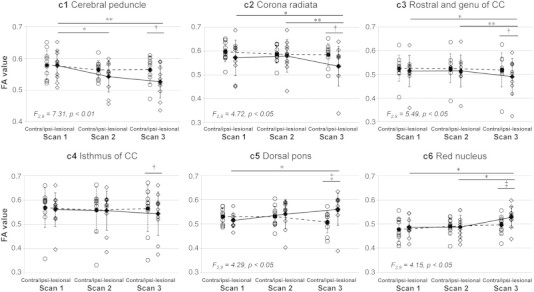
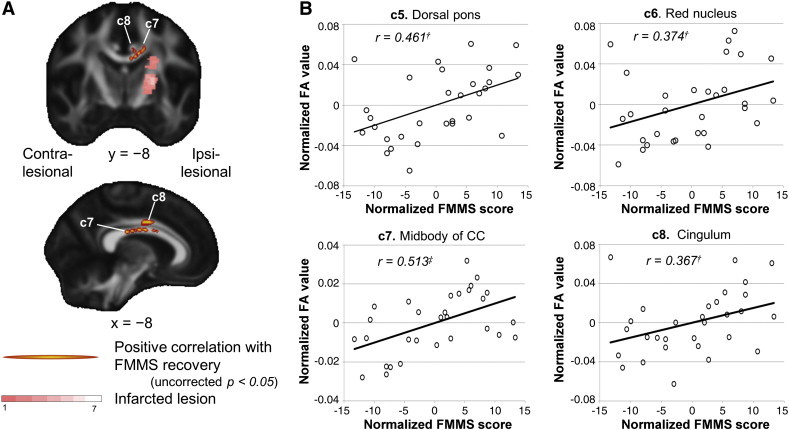
The mechanism of motor recovery after stroke may involve reorganization of the surviving networks. However, details of adaptive changes in structural connectivity are not well understood. Here, we show long-term changes in white matter microstructure that relate to motor recovery in stroke patients. We studied ten subcortical ischemic stroke patients who showed motor hemiparesis at the initial clinical examination and an infarcted lesion centered in the posterior limb of internal capsule of the unilateral hemisphere at the initial diffusion-weighted magnetic resonance imaging scan. The participants underwent serial diffusion tensor imaging and motor function assessments at three consecutive time points; within 2 weeks, and at 1 and 3 months after the onset. Fractional anisotropy (FA) was analyzed for regional differences between hemispheres and time points, as well as for correlation with motor recovery using a tract-based spatial statistics analysis. The results showed significantly increased FA in the red nucleus and dorsal pons in the ipsi-lesional side at 3 months, and significantly decreased FA in the ipsi-lesional internal capsule at all time points, and in the cerebral peduncle, corona radiata, and corpus callosum at 3 months. In the correlation analysis, FA values of clusters in the red nucleus, dorsal pons, midbody of corpus callosum, and cingulum were positively correlated with recovery of motor function. Our study suggests that changes in white matter microstructure in alternative descending motor tracts including the rubro-spinal pathway, and interhemispheric callosal connections may play a key role in compensating for motor impairment after subcortical stroke.
Keywords: CC, Corpus callosum; CP, Cerebral peduncle; CR, Corona radiata; DTI, Diffusion tensor imaging; Diffusion tensor image; EPT, Extrapyramidal tract; FA, Fractional Anisotropy; FMMS, Fugl-Meyer Motor Scale; Motor recovery; PLIC, Posterior limb of internal capsule; PT, Pyramidal tract; Reorganization; Subcortical stroke; TBSS, Tract-based spatial statistics; Tract-based spatial statistics.
Figures
Similar articles
-
Brain microstructural development at near-term age in very-low-birth-weight preterm infants: an atlas-based diffusion imaging study.Neuroimage. 2014 Feb 1;86:244-56. doi: 10.1016/j.neuroimage.2013.09.053. Epub 2013 Oct 1. Neuroimage. 2014. PMID: 24091089 Free PMC article.
-
Both projection and commissural pathways are disrupted in individuals with chronic stroke: investigating microstructural white matter correlates of motor recovery.BMC Neurosci. 2012 Aug 29;13:107. doi: 10.1186/1471-2202-13-107. BMC Neurosci. 2012. PMID: 22931454 Free PMC article.
-
Axial diffusivity changes in the motor pathway above stroke foci and functional recovery after subcortical infarction.Restor Neurol Neurosci. 2018;36(2):173-182. doi: 10.3233/RNN-170747. Restor Neurol Neurosci. 2018. PMID: 29526853
-
The role of diffusion tensor imaging and fractional anisotropy in the evaluation of patients with idiopathic normal pressure hydrocephalus: a literature review.Neurosurg Focus. 2016 Sep;41(3):E12. doi: 10.3171/2016.6.FOCUS16192. Neurosurg Focus. 2016. PMID: 27581308 Review.
-
Prediction of Upper Limb Motor Recovery after Subacute Ischemic Stroke Using Diffusion Tensor Imaging: A Systematic Review and Meta-Analysis.J Stroke. 2016 Jan;18(1):50-9. doi: 10.5853/jos.2015.01186. Epub 2016 Jan 29. J Stroke. 2016. PMID: 26846758 Free PMC article. Review.
Cited by
-
The cortico-rubral and cerebello-rubral pathways are topographically organized within the human red nucleus.Sci Rep. 2019 Aug 20;9(1):12117. doi: 10.1038/s41598-019-48164-7. Sci Rep. 2019. PMID: 31431648 Free PMC article.
-
Red nucleus structure and function: from anatomy to clinical neurosciences.Brain Struct Funct. 2021 Jan;226(1):69-91. doi: 10.1007/s00429-020-02171-x. Epub 2020 Nov 12. Brain Struct Funct. 2021. PMID: 33180142 Free PMC article. Review.
-
Restoring After Central Nervous System Injuries: Neural Mechanisms and Translational Applications of Motor Recovery.Neurosci Bull. 2022 Dec;38(12):1569-1587. doi: 10.1007/s12264-022-00959-x. Epub 2022 Nov 4. Neurosci Bull. 2022. PMID: 36333482 Free PMC article. Review.
-
Recruitment of Polysynaptic Connections Underlies Functional Recovery of a Neural Circuit after Lesion.eNeuro. 2016 Aug 26;3(4):ENEURO.0056-16.2016. doi: 10.1523/ENEURO.0056-16.2016. eCollection 2016 Jul-Aug. eNeuro. 2016. PMID: 27570828 Free PMC article.
-
Compensatory Hyperactivity of the Ipsilesional Red Nucleus in a Patient With Somatosensory Cortex Damage: A Case Report.Brain Neurorehabil. 2023 Nov 6;16(3):e33. doi: 10.12786/bn.2023.16.e33. eCollection 2023 Nov. Brain Neurorehabil. 2023. PMID: 38047094 Free PMC article.
References
-
- Beaulieu C. In: Diffusion MRI: From Quantitative Measurement to In Vivo Neuroanatomy. Johansen-Berg H., Behrens T., editors. Elsevier; London: 2009.
-
- Belhaj-Saïf A., Cheney P.D. Plasticity in the distribution of the red nucleus output to forearm muscles after unilateral lesions of the pyramidal tract. J. Neurophysiol. 2000;83:3147–3153. - PubMed
Publication types
MeSH terms
LinkOut - more resources
Full Text Sources
Other Literature Sources
Medical
Miscellaneous
