. 2014 Apr 1;459(1):71-81.
doi: 10.1042/BJ20131273.

Frataxin-bypassing Isu1: characterization of the bypass activity in cells and mitochondria

Affiliations

Frataxin-bypassing Isu1: characterization of the bypass activity in cells and mitochondria

Heeyong Yoon et al. Biochem J. .

Abstract

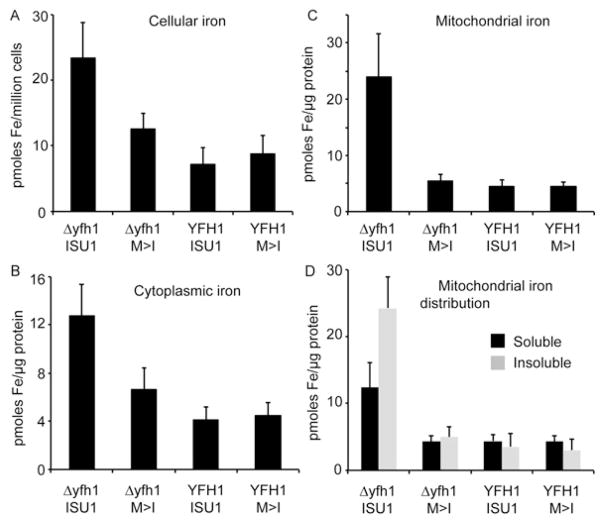
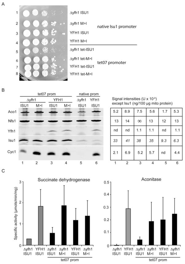
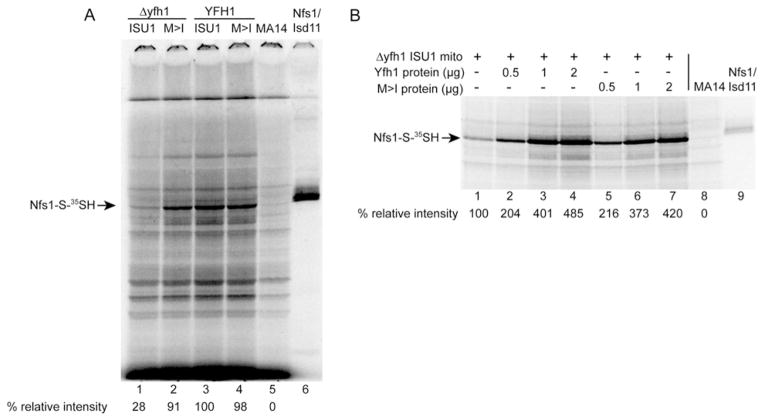
Frataxin is a conserved mitochondrial protein, and deficiency underlies the neurodegenerative disease Friedreich's ataxia. Frataxin interacts with the core machinery for Fe-S cluster assembly in mitochondria. Recently we reported that in frataxin-deleted yeast strains, a spontaneously occurring mutation in one of two genes encoding redundant Isu scaffold proteins, bypassed the mutant phenotypes. In the present study we created strains expressing a single scaffold protein, either Isu1 or the bypass mutant M107I Isu1. Our results show that in the frataxin-deletion strain expressing the bypass mutant Isu1, cell growth, Fe-S cluster protein activities, haem proteins and iron homoeostasis were restored to normal or close to normal. The bypass effects were not mediated by changes in Isu1 expression level. The persulfide-forming activity of the cysteine desulfurase was diminished in the frataxin deletion (∆yfh1 ISU1) and was improved by expression of the bypass Isu1 (∆yfh1 M107I ISU1). The addition of purified bypass M107I Isu1 protein to a ∆yfh1 lysate conferred similar enhancement of cysteine desulfurase as did frataxin, suggesting that this effect contributed to the bypass mechanism. Fe-S cluster-forming activity in isolated mitochondria was stimulated by the bypass Isu1, albeit at a lower rate. The rescuing effects of the bypass Isu1 point to ways that the core defects in Friedreich's ataxia mitochondria can be restored.


Figures

Figure 1
Figure 1. Growth of single-copy Isu strains
The four single-copy Isu strains (Δyfh1 ISU1, Δyfh1 M>I, YFH1 ISU1 and YFH1 M>I) were grown overnight in rich medium (YPAD), and 106 cells were subjected to serial dilutions and spotted on to agar plates containing 2 % glucose or 3 % ethanol as a carbon source. The plates were incubated at 30 °C for 3 days and photographed.
Figure 2
Figure 2. Enzyme activities
Mitochondria were prepared from three independent biological replicates from the four single-copy Isu strains. (A) Succinate dehydrogenase activity (means ± S.D.). Δyfh1 ISU1 was significantly different (P = 0.001) from YFH1 ISU1. YFH1 M>I was not significantly different (P = 0.422) from YFH1 ISU1. (B) Aconitase activity (means ± S.D.). Δyfh1 ISU1 was significantly different (P = 0.012) from YFH1 ISU1. YFH1 M>I was not significantly different (P = 0.368) from YFH1 ISU1. (C) The in-gel activity assay for aconitase activity on these mitochondria [39,40].
Figure 3
Figure 3. Protein levels in mitochondria
Mitochondria isolated from single-copy Isu strains were analysed by SDS/PAGE. After immunoblotting with specific rabbit antibodies for aconitase (Aco1), Nfs1, yeast frataxin homologue (Yfh1), Isu1 and Isd11, the images were developed using the LiCor system and infrared detector and the intensity signal was determined (shown in lower panel). Three independent preparations of mitochondria were evaluated, and a representative immunoblot is shown. nd, not detected.
Figure 4
Figure 4. Haem protein levels
Mitochondria isolated from single-copy Isu strains were analysed by SDS/PAGE. The proteins were transferred on to nitrocellulose membranes and probed with rabbit polyclonal antibodies against Ccp1 and Cyc1. The blots were also probed for Yfh1 and Nfs1 to confirm the genotype and to serve as controls The images were developed using the LiCor system and infrared detector and the intensity signal was determined (shown in lower panel). Three independent preparations of mitochondria were evaluated and a representative immunoblot is shown. nd, not detected.
Figure 5
Figure 5. Iron homoeostasis
The single-copy Isu strains were grown in the presence of 55Fe radionuclide tracer in defined medium supplemented with 10 μM ferrous ascorbate. Three independent labelling and fractionation experiments were performed. Results are means ± S.D. Whole cells, cytoplasmic and mitochondrial fractions were subjected to scintillation counting for 55Fe. (A) Iron per cell was determined by cell counting, and the amount in Δyfh1 ISU1 was significantly greater than the amount in YFH1 ISU1 (P = 0.010), whereas the amount in YFH1 M>I was not significantly different from the amount in YFH1 ISU1. (B) Cytoplasmic iron was determined, and the amount in Δyfh1 ISU1 was significantly greater than the amount in YFH1 ISU1 (P = 0.005), whereas the amount in YFH1 M>I was not significantly different from the amount in YFH1 ISU1. (C) Mitochondrial iron was determined and the amount in Δyfh1 was significantly greater than the amount in YFH1 ISU1 (P = 0.013), whereas the amount in YFH1 M>I was not significantly different from the amount in YFH1 ISU1. (D) For solubility determinations, mitochondria were sonicated in buffer containing 0.1 % Triton X-100. The supernatant and pellet fractions were separated by centrifugation and subjected to scintillation counting to ascertain the iron content of each fraction. The insoluble fraction was significantly greater in the Δyfh1 ISU1 than in the wt YFH1 ISU1 (P = 0.002), whereas the insoluble fraction in the YFH1 M>I was not significantly different from the amount in YFH1 ISU1.
Figure 6
Figure 6. Overexpression of Isu1 and related phenotypes
(A) Plate phenotypes. Strains with the indicated genotypes (native Isu1 promoter or tet07 promoter) were serially diluted and spotted on to YPAD agar plates. After 3 days of growth at 30 °C the plates were photographed. (B) Mitochondria were isolated from deletion (Δyfh1 ISU1) and wt (YFH1 ISU1) strains, and from the tet promoter set including Δyfh1 tet-ISU1, Δyfh1 tet-M>I, YFH1 ISU1 and YFH1 tet-M>I. Proteins were evaluated by immunoblotting using various antibodies. The images were developed using the LiCor system and infrared detector and the intensity signal was determined (shown in right-hand panel). Three independent mitochondrial preparations were analysed, and a representative immunoblot is shown. ∞, pixel saturation. Isu1 values are reported as ng/100 μg of mitochondrial protein. (C) Enzyme activities for succinate dehydrogenase (left-hand panel) and aconitase (right-hand panel) were measured in mitochondria. Three independent mitochondrial preparations were assayed. Results are means ± S.D. The mean succinate dehydrogenase activity was higher in the tet07 strain Δyfh1 tet-ISU1 compared with the native promoter strain Δyfh1 ISU1 although the difference did not reach statistical significance (P = 0.072). The mean aconitase activity was significantly higher in the tet07 strain compared with the native promoter strain (P = 0.018) as determined using a one-tailed Student’s t test.
Figure 7
Figure 7. Persulfide-forming activity in mitochondria
Mitochondria were isolated from the single-copy Isu strains (native Isu1 promoter strains). (A) Nfs1 persulfide in intact mitochondria. Intact mitochondria were depleted of endogenous nucleotides by incubation for 10 min at 30 °C. After labelling with [35S]cysteine, mitochondria were recovered, proteins were separated by non-reducing SDS/PAGE and Nfs1 persulfide (Nfs1-S-35SH) was visualized by radioautography. Lane 1, Δyfh1 ISU1; lane 2, Δyfh1 M>I; lane 3, YFH1 ISU1; lane 4, YFH1 M>I; lane 5, MA14 mitochondria; lane 6, purified Nfs1–Isd11 complex. The Nfs1 persulfide is indicated by an arrow. Four independent experiments were performed and a representative autoradiogram is shown. (B) Nfs1 persulfide in mitochondrial lysate. Lysate from the deletion (Δyfh1 ISU1) mitochondria was supplemented with different amounts of purified Yfh1 protein (lanes 2–4) or purified mutant M>I Isu1 protein (lanes 5–7). The lysates were incubated with [35S]cysteine, and radiolabelled persulfide was detected by separation on non-reducing SDS gel and radioautography. Lysate from MA14, a hypomorphic Nfs1 mutant, was included as a negative control (lane 8), and purified Nfs1–Isd11 protein complex was included as a positive control (lane 9). The experiment was repeated three times and a representative autoradiogram is shown.
Figure 8
Figure 8. Fe–S cluster forming activity in intact mitochondria
(A) Isolated mitochondria from strains with the indicated genotypes were incubated with [35S]cysteine in the presence of nucleotides (4 mM ATP, 1 mM GTP and 5 mM NADH) and ferrous ascorbate (10 μM) at 30 °C for the indicated times. Mitochondria were recovered, lysed and soluble proteins were analysed by native gel electrophoresis. Radiolabelled aconitase (Aco1 [Fe-35S]) was visualized by radioautography. The time course experiment was repeated three times and a representative autoradiogram is shown. (B) Isolated mitochondria were labelled at 30 °C for 30 min in the presence of [35S]cysteine, nucleotides and different concentrations of ferrous ascorbate (0, 10 and 50 μM). Radiolabelled aconitase was visualized as in (A). The iron titration experiment was repeated three times and a representative autoradiogram is shown.
Figure 9
Figure 9. H2 O2 sensitivity
Yeast strains (Isu1 driven by the native promoter or tet07 promoter) were serially diluted and spotted on to YPAD agar plates containing H2 O2 at different concentrations (0.25 mM, 2 mM or 4 mM). The growth of the bypass strain (Δyfh1 M>I) was indistinguishable from the wt strain (YFH1 ISU1) at low H2 O2, but severely impaired at high H2 O2 concentration.

Similar articles

Cited by

References

    1. Campuzano V, Montermini L, Molto MD, Pianese L, Cossee M, Cavalcanti F, Monros E, Rodius F, Duclos F, Monticelli A, et al. Friedreich’s ataxia: autosomal recessive disease caused by an intronic GAA triplet repeat expansion. Science. 1996;271:1423–1427. - PubMed
    1. Rouault TA. Biogenesis of iron–sulfur clusters in mammalian cells: new insights and relevance to human disease. Dis Model Mech. 2012;5:155–164. - PMC - PubMed
    1. Whitnall M, Suryo Rahmanto Y, Huang ML, Saletta F, Lok HC, Gutierrez L, Lazaro FJ, Fleming AJ, St Pierre TG, Mikhael MR, et al. Identification of nonferritin mitochondrial iron deposits in a mouse model of Friedreich ataxia. Proc Natl Acad Sci USA. 2012;109:20590–20595. - PMC - PubMed
    1. Wilson RB, Roof DM. Respiratory deficiency due to loss of mitochondrial DNA in yeast lacking the frataxin homologue. Nat Genet. 1997;16:352–357. - PubMed
    1. Yoon H, Golla R, Lesuisse E, Pain J, Donald JE, Lyver ER, Pain D, Dancis A. Mutation in the Fe–S scaffold protein Isu bypasses frataxin deletion. Biochem J. 2012;441:473–480. - PMC - PubMed

Publication types

MeSH terms