. 2014 Jan 17:15:15.
doi: 10.1186/1471-2202-15-15.

Morphological changes of cortical pyramidal neurons in hepatic encephalopathy

Affiliations

Morphological changes of cortical pyramidal neurons in hepatic encephalopathy

Jeng-Rung Chen et al. BMC Neurosci. .

Abstract

Background: Hepatic encephalopathy (HE) is a reversible neuropsychiatric syndrome associated with acute and chronic liver diseases. It includes a number of neuropsychiatric disturbances including impaired motor activity and coordination, intellectual and cognitive function.

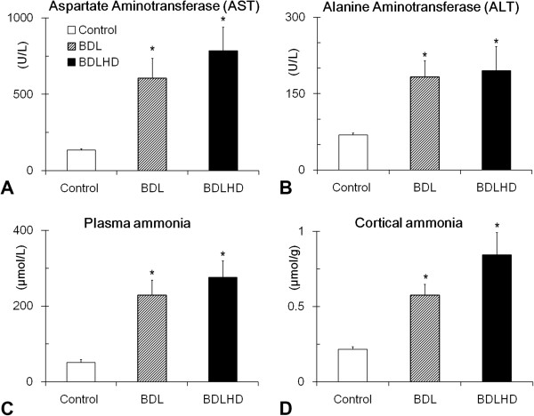
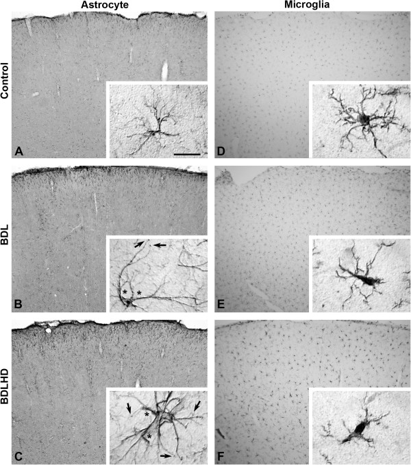
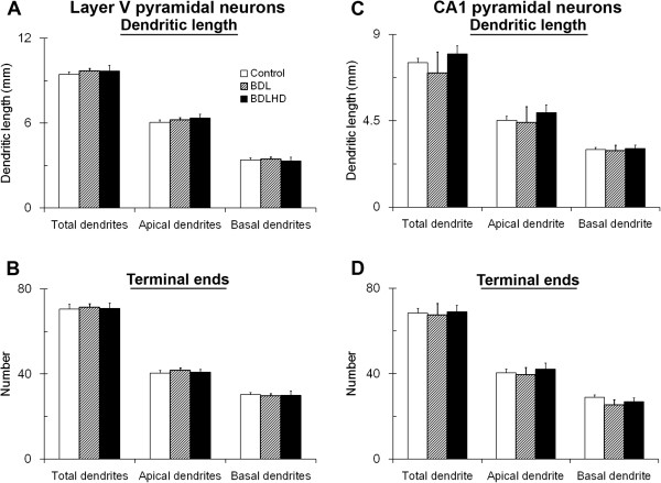
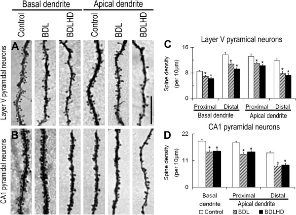
Results: In the present study, we used a chronic rat HE model by ligation of the bile duct (BDL) for 4 weeks. These rats showed increased plasma ammonia level, bile duct hyperplasia and impaired spatial learning memory and motor coordination when tested with Rota-rod and Morris water maze tests, respectively. By immunohistochemistry, the cerebral cortex showed swelling of astrocytes and microglia activation. To gain a better understanding of the effect of HE on the brain, the dendritic arbors of layer V cortical pyramidal neurons and hippocampal CA1 pyramidal neurons were revealed by an intracellular dye injection combined with a 3-dimensional reconstruction. Although the dendritic arbors remained unaltered, the dendritic spine density on these neurons was significantly reduced. It was suggested that the reduction of dendritic spines may be the underlying cause for increased motor evoked potential threshold and prolonged central motor conduction time in clinical finding in cirrhosis.

Conclusions: We found that HE perturbs CNS functions by altering the dendritic morphology of cortical and hippocampal pyramidal neurons, which may be the underlying cause for the motor and intellectual impairments associated with HE patients.


Figures

Figure 1
Figure 1
Representative micrographs showing the liver lobules in HE rat. Bar = 100 μm in A-C.
Figure 2
Figure 2
Biochemical values in HE rats. The AST (A), ALT (B) and ammonia levels of serum (C) and cerebral cortex (D) or HE rats were measured. *, p < 0.05 between the experimental rats and sham control.
Figure 3
Figure 3
Behavioral performances in HE rats. The Rotarod (A) and Morris water maze (B, C and D) tests were analyzed in BDL and BDLHD rats. These data was normalized to those obtained before HE induction. *, p < 0.05 between the marked and control rat; #, p < 0.05 between the marked and BDL rats.
Figure 4
Figure 4
Representative micrographs showing astrocytes and microglia in HE rats. The left column was GFAP-immunoreactive astrocytes (A-C) and right column was Iba1-immunoreactive microglia (D-F) in sham control (A and D), BDL (B and E) and BDLHD (C and F). At a high magnification, thickened processes (*) and bouton-like terminals (arrow) were easily identified in BDL and BDLHD rats. The polyclonal Iba1 antibodies labeled both inactive microglia (insert of D) and activated microglia (inserts of E and F). Bar = 300 μm in A-C, 20 μm for inserts.
Figure 5
Figure 5
The cytoarchitecture of sensorimotor cortex in HE rats. The soma size of astrocytes and layer III / V pyramidal neurons (A and B) and density of nNOS+ neurons (C) and microglia (D) were measured in sensorimotor cortex. *, p < 0.05 between the mark and control rats.
Figure 6
Figure 6
Representative 3-dimensionally reconstructed pyramidal neurons. The layer V (A – C) pyramidal neurons of the primary sensorimotor cortex and hippocampal CA1 (D – F) pyramidal neurons were reconstructed with Neurolucida®. The apical dendritic trunk was in red while the filled blue circle represents cell body. Branches of each basal dendritic trunk were displayed with the same color. Roman numerals and horizontal bars on the left of each drawing mark the cortical layers. Bar = 200 μm in A-C and 100 μm in D-F.
Figure 7
Figure 7
The dendritic arbor of pyramidal neurons was analyzed in HE rats. The dendritic arbors of layer V (A and B) pyramidal neurons of the primary sensorimotor cortex and hippocampal CA1 (C and D) pyramidal neurons were analyzed.
Figure 8
Figure 8
The spine loss of pyramidal neurons in HE rats. The spine density on layer V (A) pyramidal neurons of the primary sensorimotor cortex and hippocampal CA1 (B) pyramidal neurons was analyzed. Representative micrographs of basal segment and distal apical segment of dendrite were illustrated in A and B. Spine density was measured and analyzed in C and D. *, p < 0.05 between the mark and control rats. Bar = 10 μm for all photograph.

Similar articles

Cited by

References

    1. Patel D, McPhail MJ, Cobbold JF, Taylor-Robinson SD. Hepatic encephalopathy. Br J Hosp Med (Lond) 2012;15(2):79–85. - PubMed
    1. Bosoi CR, Rose CF. Identifying the direct effects of ammonia on the brain. Metab Brain Dis. 2009;15(1):95–102. doi: 10.1007/s11011-008-9112-7. - DOI - PubMed
    1. Romero-Gomez M, Jover M, Galan JJ, Ruiz A. Gut ammonia production and its modulation. Metab Brain Dis. 2009;15(1):147–157. doi: 10.1007/s11011-008-9124-3. - DOI - PubMed
    1. Walker V. Severe hyperammonaemia in adults not explained by liver disease. Ann Clin Biochem. 2012;15(Pt 3):214–228. - PubMed
    1. Rama Rao KV, Norenberg MD. Glutamine in the pathogenesis of hepatic encephalopathy: the trojan horse hypothesis revisited. Neurochem Res. DOI 10.1007/s11064-012-0955-2. - PMC - PubMed

Publication types

LinkOut - more resources