. 2014 Jan 16:14:28.
doi: 10.1186/1471-2407-14-28.

STMN-1 is a potential marker of lymph node metastasis in distal esophageal adenocarcinomas and silencing its expression can reverse malignant phenotype of tumor cells

Affiliations

STMN-1 is a potential marker of lymph node metastasis in distal esophageal adenocarcinomas and silencing its expression can reverse malignant phenotype of tumor cells

Javed Akhtar et al. BMC Cancer. .

Retraction in

Abstract

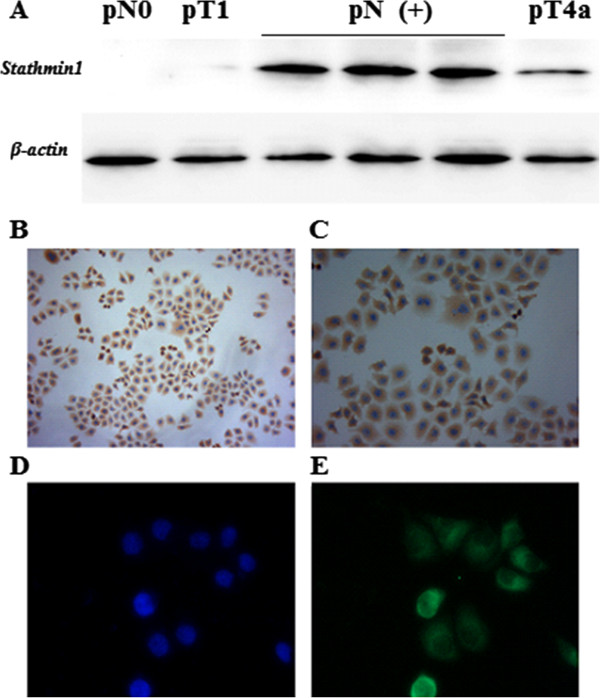
Background: Distal esophageal adenocarcinoma is a highly aggressive neoplasm. Despite advances in diagnosis and therapy, the prognosis is still poor. Stathmin (STMN-1) is a ubiquitously expressed microtubule destabilizing phosphoprotein. It promotes the disassembly of microtubules and prevents assembly. STMN-1 can cause uncontrolled cell proliferation when mutated and not functioning properly. Recently, found to be overexpressed in many types of human cancers. However, its clinical significance remains elusive in distal esophageal adenocarcinoma. Here, we reported for the first time that STMN-1 is highly overexpressed in adenocarcinomas of the distal esophagus and strongly associated with lymph node metastasis.

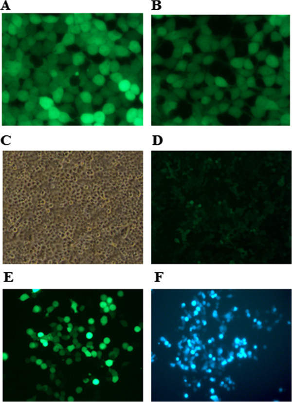
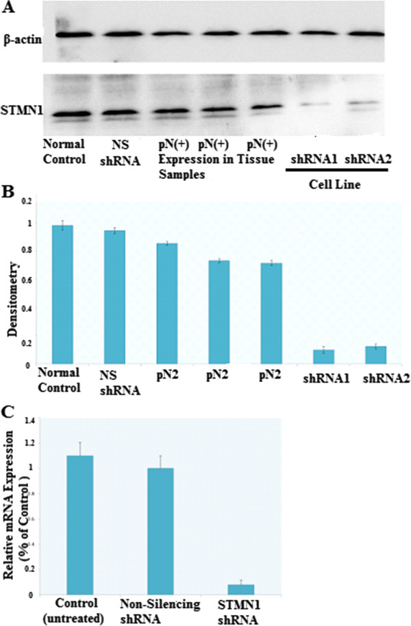
Methods: STMN-1 expression in 63 cases of distal esophageal adenocarcinoma was analyzed by immunoblotting, while expression in esophageal adenocarcinoma cells was determined by immunocytochemistry, immunofluorescence, qRT-PCR and western blotting. Lentivirus-mediated RNAi was employed to knock-down STMN-1 expression in Human esophageal adenocarcinoma cells. The relationship between STMN-1 expression and lymph node metastasis in distal esophageal adenocarcinoma was determined by univariate and multivariate analyses.

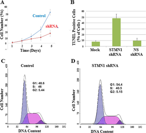
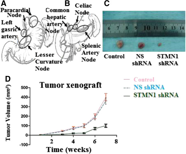
Results: STMN-1 was detected in 31 (49.21%) of the 63 cases. STMN-1 was highly overexpressed in specimens with lymph node metastasis pN (+), but its expression was almost undetected in pN (-) status. Multivarian regression analysis demonstrated that STMN-1 overexpression is an independent factor for lymph node metastasis in distal esophageal adenocarcinoma. STMN-1 shRNA effectively reduced STMN-1 expression in esophageal adenocarcinoma cells (P < 0.05), which significantly suppressed proliferation (P < 0.05), increased migration (P < 0.05) and invasion ability (P < 0.05) and G1 phase arrest (P < 0.05) which lead to induction of apoptosis in esophageal adenocarcinoma cells in vitro. To verify the in vitro data, we conducted in vivo tumor xenograft studies. Esophageal adenocarcinoma cells stably transfected with STMN-1 shRNA significantly reduced tumor xenografts volume in vivo.

Conclusions: STMN-1 overexpression is associated with lymph node metastasis and increase malignancy in distal esophageal adenocarcinoma. In vivo and in vitro laboratory findings, suggests that STMN-1 may be a suitable target for future therapeutic strategies in distal esophageal adenocarcinoma.


Figures

Figure 1
Figure 1
STMN1 expression in the distal esophageal adenocarcinoma tissue samples and cell lines. (A) Distal esophageal adenocarcinoma tissue samples were lysed and immunoblotting was performed to detect expression of STMN1. Actin was used as loading control. STMN1 expression was low or negative in the samples obtained from the patients without lymph node metastasis (pN0) and pT1 classification but in the patients with lymph node metastasis (pN+) status STMN1 was highly Overexpressed, some pT4a stage patients also showed moderate STMN1 expression. (B) Light microscopic immunocytochemistry revealed an intense cytoplasmic presence of STMN1 protein in esophageal cancer cells. (C) Image at High-resolution showed strong cytoplasmic expression of STMN1. (D) Fluorescence microscopy of DAPI stained cells. (E) Immunofluorescent images of distal esophageal adenocarcinoma cells labeled with an antibody against STMN1, the staining was repeated in triplicate three independent times.
Figure 2
Figure 2
Transfection, Lenti-virus production and transduction. Co-transfection of the Trans-Lentiviral packaging mix with a shRNA transfer vector into HEK293T packaging cells was done using Arrest-In Transfection Reagent. Following co-transfection, replication-incompetent virions were released into the media which were collected after 48 h and 72 respectively. (A) Fluorescence microscopy image showing TurboGFP expression from the pGIPZ STMN1 vector cells 72 h post-transfection. (B) Image showing the TurboGFP expression from the GIPZ Non-silencing Control. (C) Phase contrast microscopy image. (D) TurboGFP images showing shRNA delivery efficiency. (E) &(F) post puromycine selection images showing efficient transduction of Lenti-STMN1 shRNA in distal esophageal cancer cells.
Figure 3
Figure 3
Verification of knockdown of STMN1 gene expression in esophageal cancer cell line by lentivirus-mediated RNA interference. (A) Western blot analysis of STMN1 protein in lysates of either untreated (control) or transfected with a Non-silencing shRNA (scrambled sequence) and transfected with a specific STMN1 shRNA. ACTIN expression was used as a loading control. (B) Densitometry analysis of A normalized to Actin, Laser densitometric analysis of protein bands was performed, and the ratio of stathmin expression to Beta-Actin expression (STMN:Beta-Actin) was determined and normalized. (C) Total RNA isolated from distal esophageal adenocarcinoma cells before and after silencing stathmin gene expression using STMN1 shRNA were analyzed with real-time RT–PCR. There was no significant difference between controls and non-silencing shRNA (scrambled sequences). Data are expressed as percentage change (Means ± S.D.) compared with controls and represent four independent experiments. (P < 0.05 vs Non-silencing shRNA, one-way analysis of variance (ANOVA) followed by Tukey’s multiple comparion).
Figure 4
Figure 4
Effects of STMN1 knockdown on cell growth and apoptosis. (A) Cells were transfected with Non-silencing shRNA as a negative control or STMN1shRNA. Cell proliferation was measured by CCK-8. Data are expressed as percentage change (Means ± S.D.) compared with controls and represent six independent experiments. (P < 0.05 vs Non-silencing shRNA, one-way analysis of variance (ANOVA) followed by Tukey’s multiple comparion). (B) Apoptosis was assessed by TUNEL assay, the amount of DNA fragmentation (apoptosis) was assessed by TUNEL assay for esophageal cancer cells before and after silencing stathmin gene expression utilizing STMN1 shRNA. There was no significant difference between controls and non-silencing shRNA (scrambled sequences). Experiments were performed in triplicate. (C) Cell-cycle distributions of control shRNA infected cells and (D) STMN1 shRNA infected cells as measured by flow cytometry.
Figure 5
Figure 5
Effects of STMN1 knockdown on cell migration and invasion. The images of cells migrating PVPF filters as examined by cell migration assay using Boyden chambers. Cell migration was evaluated in the Boyden migration assay two days after Esophageal cancer cells were either (A) untreated or (B) transfected with Non-silencing shRNA or (C) stathmin1 siRNA. (D) Representing average of STMN1 shRNA infected cells, Non-silencing shRNA infected cells and non-infected cells (P < 0.05) versus control. (E) The average invading cell counts of STMN1 shRNA infected cells, Non-silencing shRNA infected cells and non-infected cells. Data are expressed as percentage change (Means ± S.D.) compared with controls and represent four independent experiments. (P < 0.05 vs Non-silencing shRNA, one-way analysis of variance (ANOVA) followed by Tukey’s multiple comparion).
Figure 6
Figure 6
Abdominal lymph node clearance and xenograft tumor models in nude mice. (A) Perigastric lymph nodes, lymph nodes along the left gastric artery and lesser curvature lymph nodes dissection. (B) Distant abdominal lymph node dissection. Abdominal Esophagus drains into superior gastric artery, celiac axis, common hepatic artery and splenic artery lymph nodes. (C) Esophageal cancer cells were either untreated or transfected with Non-silencing shRNA (scrambled sequence) as a negative control and transfected with STMN1 shRNA were xenografted subcutaneously in the BALB/c-nu/nu male mice. Tumor mass (xenograft) volume was measured every week from week 3 to week 7. Data are expressed as percentage change (Means ± S.D.) compared with controls and represent four independent experiments. (P < 0.05 vs Non-silencing shRNA, one-way analysis of variance (ANOVA) followed by Tukey’s multiple comparion). (D) Photograph of xenografts dissected from nude mice after 7 weeks subcutaneous inoculation showing suppression growth of cancer cells transfected with stathmin1 shRNA as compared to cells transfected with untreated or transfected with Non-silencing shRNA.

References

    1. Jemal A, Siegel R, Xu J, Ward E. Cancer statistics. CA Cancer J Clin. 2010;60:277–300. doi: 10.3322/caac.20073. - DOI - PubMed
    1. Jemal A, Bray F, Center MM, Ferlay J, Ward E, Forman D. Global cancer statistics. CA Cancer J Clin. 2011;61:69–90. doi: 10.3322/caac.20107. - DOI - PubMed
    1. Kachroo S, Etzel CJ. Decreasing the cancer burden in developing countries: concerns and recommendations. Eur J Cancer Care (Engl) 2009;18:18–21. doi: 10.1111/j.1365-2354.2008.00985.x. - DOI - PubMed
    1. Sobin LH, Gospodarowicz MK, Wittekind C. TNM Classification of Malignant Tumours. 7. Oxford, UK: Wiley-Blackwell; 2009.
    1. AJCC Cancer Staging Handbook. ftACSM. 7. New York, NY: Springer; 2010. pp. 129–143.

Publication types

MeSH terms

Supplementary concepts