Review
. 2014 Jun;133(6):1521-34.
doi: 10.1016/j.jaci.2013.11.027. Epub 2014 Jan 13.

Mechanisms underlying the neuronal-based symptoms of allergy

Affiliations
Review

Mechanisms underlying the neuronal-based symptoms of allergy

Bradley J Undem et al. J Allergy Clin Immunol. 2014 Jun.

Abstract

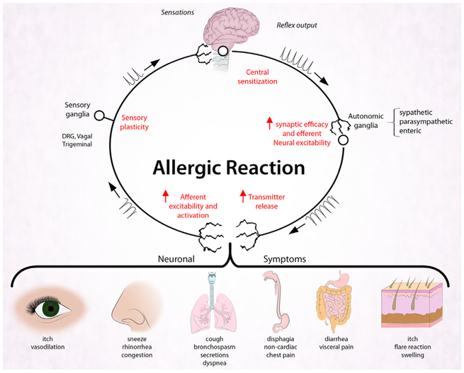
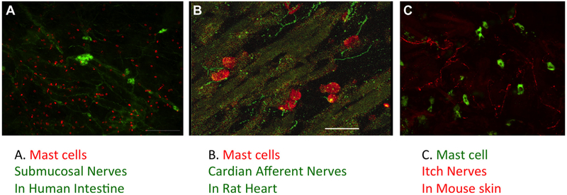
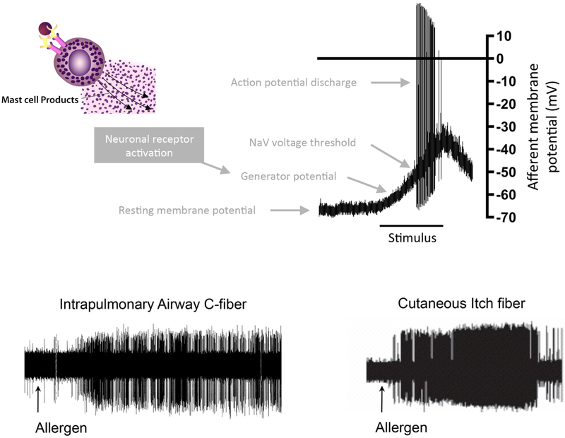
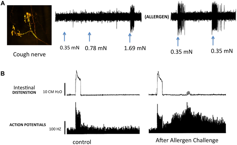
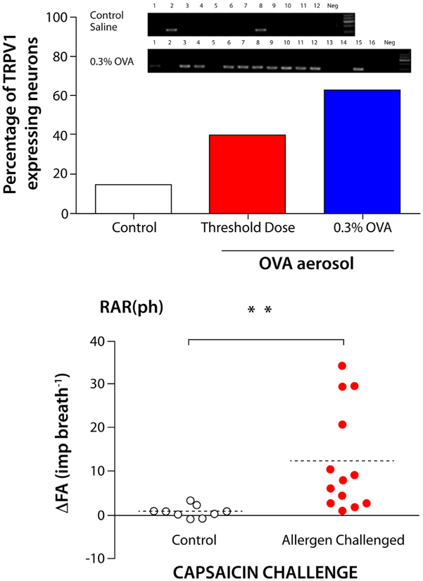
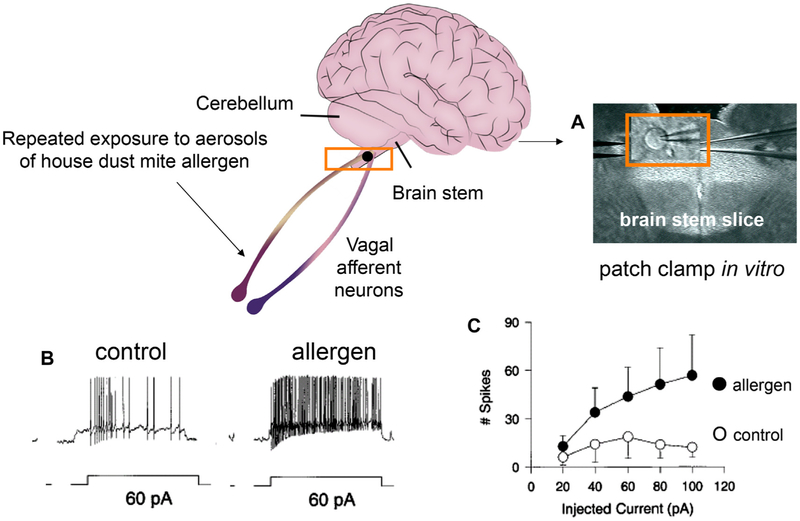
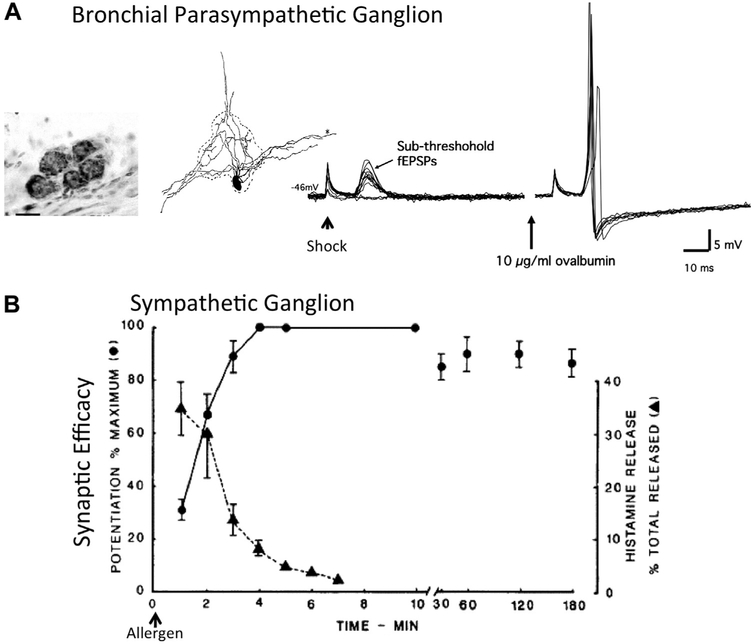
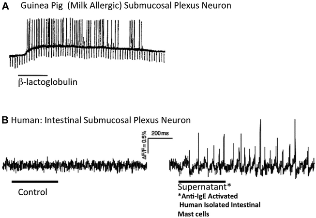
Persons with allergies present with symptoms that often are the result of alterations in the nervous system. Neuronally based symptoms depend on the organ in which the allergic reaction occurs but can include red itchy eyes, sneezing, nasal congestion, rhinorrhea, coughing, bronchoconstriction, airway mucus secretion, dysphagia, altered gastrointestinal motility, and itchy swollen skin. These symptoms occur because mediators released during an allergic reaction can interact with sensory nerves, change processing in the central nervous system, and alter transmission in sympathetic, parasympathetic, and enteric autonomic nerves. In addition, evidence supports the idea that in some subjects this neuromodulation is, for reasons poorly understood, upregulated such that the same degree of nerve stimulus causes a larger effect than seen in healthy subjects. There are distinctions in the mechanisms and nerve types involved in allergen-induced neuromodulation among different organ systems, but general principles have emerged. The products of activated mast cells, other inflammatory cells, and resident cells can overtly stimulate nerve endings, cause long-lasting changes in neuronal excitability, increase synaptic efficacy, and also change gene expression in nerves, resulting in phenotypically altered neurons. A better understanding of these processes might lead to novel therapeutic strategies aimed at limiting the suffering of those with allergies.

Keywords: Allergy; C-fiber; IgE; action potential; afferent; airways; allergen; anti-inflammatory; autonomic; bronchospasm; cough; critical periods; cytokine; degranulation; depolarization; dorsal root; dysfunction; efferent; enteric; eye; ganglia; gut; histamine; hypersecretion; inflammation; innervation; ion channel; irritant; itch; jugular; leukotriene; lungs; mast cell; mast cell mediators; mast cell-nerve interactions; motility; myenteric; nerve; nociceptor; nodose; nucleus tractus solitarious; pain; parasympathetic; phenotypic switch; plasticity; prostaglandin; receptor; reflex; sensory; skin; steroids; sympathetic; symptoms; transient receptor potential channels; trigeminal; vagal.


Conflict of interest statement

Disclosure of potential conflict of interest: B. Undem has received grants from the National Institutes of Health and GlaxoSmithKline; is on the scientific advisory board for Afferent Pharmaceuticals; and has consultant arrangements with GlaxoSmithKline. T. Taylor-Clark declares that he has no relevant conflicts of interest.

Figures

FIG 1.
FIG 1.
Neuromodulation during the allergic reaction. Experimental studies in vivo and ex vivo support the hypothesis that the allergenic response can involve neuromodulation along the sensory (afferent)–CNS–autonomic/enteric nerve axis (see text for details). The neuromodulation ultimately leads to many of the symptoms of allergic disease. DRG, Dorsal root ganglion.
FIG 2.
FIG 2.
Mast cells are found in close proximity to nerves in virtually all organs. A, Mast cell tryptase–positive cells (red) near PGP9.5-positive nerves (green) in human intestinal submucosal plexus. B, Mast cells (red) near synapsin-positive neurons (green) in rat cardiac ventricle. C, Mast cells (purple) near MrgA3 expressing “afferent itch nerves” (orange) in mouse skin (personal observation).
FIG 3.
FIG 3.
Allergen challenge overtly activates afferent C-fibers. Top, Hypothetical effect of allergenic activation of mast cells on afferent nerve terminal action potential discharge. Bottom, Examples of allergen-induced activation of afferent nociceptors: left, allergen (ovalbumin) evokes strong activation of vagal jugular C-fiber innervating the lung (action potentials recorded in vagal sensory ganglion); right, allergen (mosquito extract) induces the activation of a somatosensory itch fiber in the skin (action potentials recorded in the dorsal horn).
FIG 4.
FIG 4.
Allergen-induced increase in sensory nerve excitability. A, Nodose Aδ “cough” fiber terminals in the guinea pig trachea; increased sensitivity to mechanical stimulation of this fiber type after allergen (ovalbumin) challenge. B, Multiunit recording of intestinal afferent nerves demonstrating markedly enhanced (prolonged) response to distension after allergen (ovalbumin) challenge.
FIG 5.
FIG 5.
Examples of allergen-induced sensory neuroplasticity. Top, RT-PCR from individual neurons retrogradely traced from the trachea. The example shows 12 neurons from control-treated guinea pigs and 16 neurons from ovalbumin (OVA)–treated guinea pigs. The histograms show the percentage of trachea-specific nodose neurons in each treatment group that express TRPV1 mRNA. Allergen (ovalbumin) challenge induces de novo expression of TRPV1 in nodose Aδ “cough” fibers innervating the trachea (from Lieu et al). Bottom, Allergen challenge induces de novo capsaicin sensitivity in lung rapidly adapting receptor (RAR) fibers that have been characterized by their phasic response during respiration (RARph). ΔFA, Difference in firing activity. Used with permission from Zhang et al. **P < .01.
FIG 6.
FIG 6.
Allergen-induced increases in electrical excitability in CNS neurons. Top, Brainstem location of the nucleus of the solitary tract. A, Photomicrograph of a patch-clamped neuron in a brain slice from the caudomedial nucleus of the solitary tract, which is where the vagal sensory afferents terminate. B, Whole-cell patch-clamp recordings of depolarizing current pulses applied to individual nucleus of the solitary tract neurons from naive or allergen-challenged (dust mite) rhesus monkeys. C, Increased action potential discharge in response to a depolarizing stimulus in neurons isolated from allergic monkeys.
FIG 7.
FIG 7.
Allergen-induced increases in synaptic efficacy in autonomic ganglia. A, Small parasympathetic bronchial ganglion in the guinea pig bronchus. A single neuron filled with horseradish peroxidase and drawn by using the camera lucida technique is shown. The parasympathetic neuron’s synaptic efficacy in response to stimulation of preganglionic nerves (shock artifact) is greatly increased after allergen (ovalbumin) exposure. fEPSPs, Fast excitatory postsynaptic potentials. B, Time course of histamine release and superior cervical ganglia synaptic efficacy (postganglionic compound action potential magnitude) during allergen (ovalbumin) challenge.
FIG 8.
FIG 8.
Allergen-induced stimulation of enteric neurons. A, Intracellular recording of a neuron in the submucosal plexus isolated from a guinea pig shows neuronal depolarization to action potential threshold after milk allergen challenge. B, Action potential recording from a neuron in the submucosal plexus of the human intestine. Action potentials were evoked in response to treatment with the supernatant solution of anti-IgE–activated human intestinal mast cells. Used with permission from Schemann et al.

References

    1. Lynn B Somatosensory receptors and their CNS connections. Annu Rev Physiol 1975;37:105–27. - PubMed
    1. Sherrington C The integrative action of the nervous system. New Haven (CT): Yale University Press; 1906.
    1. Coleridge JC, Coleridge HM. Afferent vagal C fibre innervation of the lungs and airways and its functional significance. Rev Physiol Biochem Pharmacol 1984; 99:1–110. - PubMed
    1. Wood JD. Enteric neuroimmunophysiology and pathophysiology. Gastroenterology 2004;127:635–57. - PubMed
    1. Blackshaw LA, Brookes SJ, Grundy D, Schemann M. Sensory transmission in the gastrointestinal tract. Neurogastroenterol Motil 2007;19:1–19. - PubMed

Publication types