. 2014 Jan 13;25(1):21-36.
doi: 10.1016/j.ccr.2013.12.007.

CARM1 methylates chromatin remodeling factor BAF155 to enhance tumor progression and metastasis

Affiliations

CARM1 methylates chromatin remodeling factor BAF155 to enhance tumor progression and metastasis

Lu Wang et al. Cancer Cell. .

Erratum in

Abstract

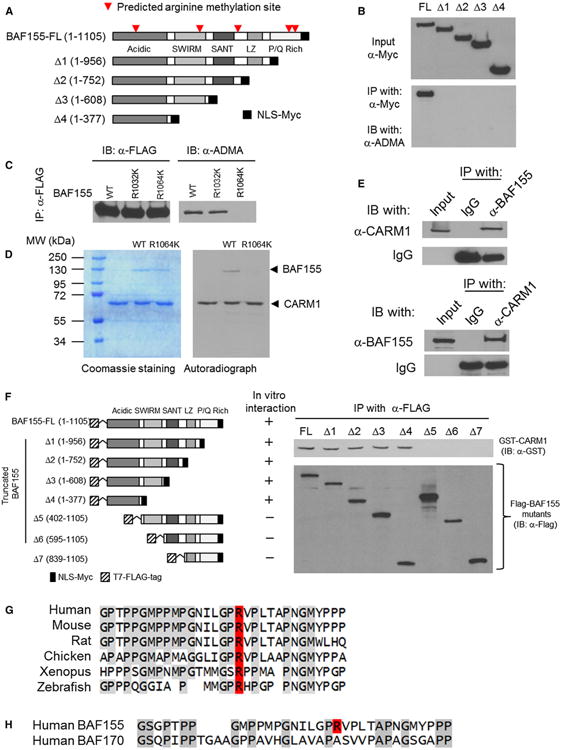
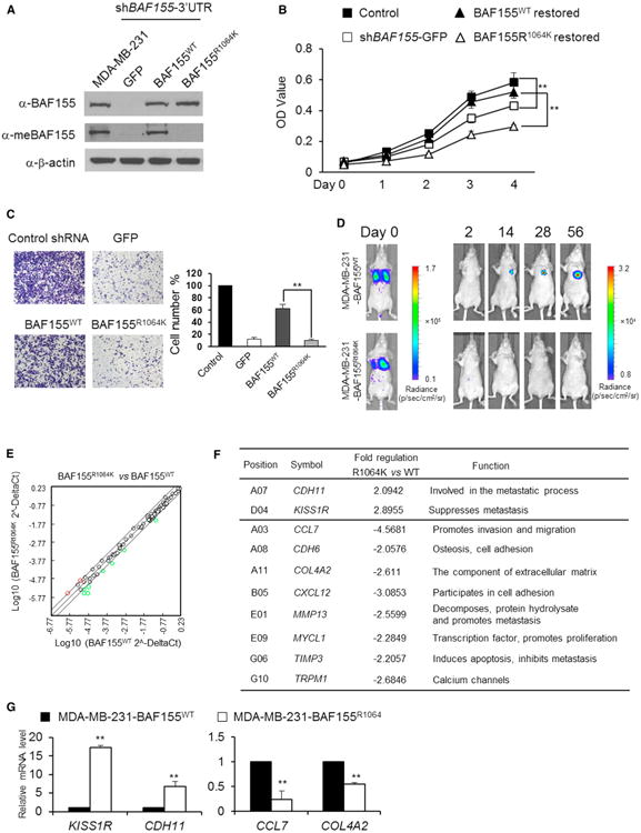
Coactivator-associated arginine methyltransferase 1 (CARM1), a coactivator for various cancer-relevant transcription factors, is overexpressed in breast cancer. To elucidate the functions of CARM1 in tumorigenesis, we knocked out CARM1 from several breast cancer cell lines using Zinc-Finger Nuclease technology, which resulted in drastic phenotypic and biochemical changes. The CARM1 KO cell lines enabled identification of CARM1 substrates, notably the SWI/SNF core subunit BAF155. Methylation of BAF155 at R1064 was found to be an independent prognostic biomarker for cancer recurrence and to regulate breast cancer cell migration and metastasis. Furthermore, CARM1-mediated BAF155 methylation affects gene expression by directing methylated BAF155 to unique chromatin regions (e.g., c-Myc pathway genes). Collectively, our studies uncover a mechanism by which BAF155 acquires tumorigenic functions via arginine methylation.


Figures

Figure 1
Figure 1. Generation of CARM1 KO Breast Cancer Cell Lines
(A) Western blot analysis of CARM1 and methyl-PABP1 using whole cell lysates from CARM1+/+ MEF, CARM1−/− MEF, MCF7, and MCF7-shCARM1 cells. β-Actin was used as a loading control. (B) Targeting exon 8 of human CARM1 by ZFN. (C) Western blot of CARM1, PABP1, methyl-PABP1, PRMT5 in parental and CARM1 KO MCF7, MDA-MB-231, and HEK293T cells. (D) Genomic DNA sequence of ZFN targeting site in MCF7 CARM1 KO and MDA-MB-231 CARM1 KO cell clones. (E) ZFN-mediated KO frequency of CARM1 in each indicated cell line. (F) Western blot of CARM1, PABP1, and me-PABP1 in MCF7, two MCF7 CARM1 KO clones, and MCF7 CARM1 KO clones re-expressing CARM1 by retroviral infection. (G) Representative cell morphology images of MCF7 cells, MCF7-shCARM1 cells, and the MCF7-derived CARM1 KO cell lines listed in (F). Scale bar = 40 μm. See also Figure S1.
Figure 2
Figure 2. Identification of BAF155 as a Substrate for CARM1
(A) Western blot analysis of total proteins in MCF7 and MCF7 CARM1 KO cells using antibodies against MMA and ADMA moieties. (B) Coomassie brilliant blue staining of proteins immunoprecipitated with anti-ADMA antibody from total cell lysates of MCF7 and MCF7 CARM1 KO cells. Discrete bands in the high molecular weight range were excised and subjected to mass spectrometry analysis, from which ten candidate proteins were identified (right panel). (C) Western blot analysis of input and anti-ADMA immunoprecipitated proteins using antibodies to detect endogenous BRG1, BAF170, BAF155, and BAF250, or against the FLAG epitope to detect transfected, FLAG-tagged TRAP150, Caprin1, TRAP230, and BCLAF1 in HEK293T and HEK293T CARM1 KO cells. (D) Coomassie brilliant blue staining (left panel) and autoradiograph (right panel) of in vitro methylated BAF155 by CARM1 in the presence of 3H-SAM. (E) Western blot analysis of anti-BAF155 immunoprecipitated proteins using anti-ADMA, BAF155, and CARM1 antibodies in HEK293T CARM1 KO cells before and after restoration with WT CARM1 or a methylation-defective CARM1 mutant (MUT). See also Figure S2.
Figure 3
Figure 3. CARM1 Methylates BAF155 at a Single Site, R1064
(A) Schematic diagram of BAF155 full-length cDNA and C-terminally truncated derivatives, each with a nuclear localization sequence/Myc (NLS-Myc) epitope tag cassette fused to its C terminus. Red arrows point to putative arginine methylation sites predicted by the PMeS program. (B) Plasmids expressing the BAF155 cDNA derivatives shown in (A) were transiently transfected into HEK293 cells and detected by western blotting with anti-Myc antibody (upper panel). Total cell lysates were immunoprecipitated with anti-Myc antibody and western blotted with anti-ADMA antibody (lower panel). (C) FLAG-tagged recombinant BAF155WT, BAF155R1032K, and BAF155R1064K proteins were immunoprecipitated from HEK293T cells with anti-FLAG antibody and detected by western blotting using anti-FLAG (left panel) or anti-ADMA (right panel) antibodies. (D) In vitro assay using recombinant CARM1 and 3H-SAM to test for methylation of BAF155WT and BAF155R1064K proteins prepared from HEK293T CARM1 KO cells. (E) Reciprocal coimmunoprecipitation of CARM1 with BAF155, and BAF155 with CARM1, from MCF7 cell lysates. (F) Mapping of CARM1-interacting domain to the N-terminal acidic domain of BAF155 by GST-pull-down assay. Left panel shows schematic diagrams of FLAG-tagged BAF155 truncation derivatives that were incubated with GST-CARM1 proteins and immunoprecipitated with anti-FLAG antibody. The immunoprecipitates were tested for the presence of GST-CARM1 by western blotting using anti-GST antibody (right panel). (G) CLUSTALW alignment of vertebral BAF155 sequences flanking human BAF155 methylation site R1064, which is highlighted in red. (H) CLUSTALW alignment of P/Q-rich domain sequences of BAF155 (with R1064 highlighted in red) and its homolog BAF170 protein. See also Figure S3 and Table S1.
Figure 4
Figure 4. BAF155 Methylation Promotes MCF7 Cell Colony Formation and Directs Unique Genomic Association Patterns
(A) Western blotting of BAF155 and me-BAF155 in MCF7 cells and MCF7-shBAF155 cells restored with GFP, BAF155WT, or BAF155R1064K. (B) Representative images of cell morphology (upper panel; scale bars = 40 μm) and colonies (lower panel; scale bars = 100 μm) for parental MCF7 and MCF7-shBAF155-GFP cells, and BAF155WT- and BAF155R1064K-restored MCF7 cells. (C) Colony yields for the cell lines in (B). (D) Western blotting analysis of CARM1, BAF155, and me-BAF155 proteins in MCF7 CARM1 KO clone 1, MCF7-CARM1KO-BAF155WT, and MCF7-CARM1KO-BAF155R1064K cells engineered to express either GFP or CARM1. β-Actin was used as loading control. (E) Representative images of colonies formed by each cell line shown in (D). Scale bar = 100 mm. (F) Colony yields for the cell lines in (D). (G) ChIP-seq identifies BRG1, BAF155WT, and BAF155R1064K association in an ∼750 kb region of chromosome 8. The coordinates shown are in hg19 and all regions were identified in MCF7and HeLa cells as detailed in Table S2. An “a” denotes a peak that is detected by BRG1 and BAF155 in both HeLa and MCF7; “b” denotes BAF155 binding peak only found in HeLa cells; “c” denotes shared BAF155and BRG1 binding peaks in HeLa but not in MCF7 cells; “d” denotes shared BAF155WT and BAF155R1064K peaks only found in MCF7 cells; and “e” denotes BAF155WT binding only found in MCF7 cells. (H) Venn diagram showing the overlapping and discrete BAF155cistromes in MCF7 cells expressing BAF155WTand BAF155R1064K. Genomic-region-associated genes were defined as the regions between 5.0 kb upstream and 1.0 kb downstream of annotated genes, using GREAT. (I) Distribution of the genome-wide association of BAF155 binding sites in MCF7 cells is comparable to that in HeLa cells. (J) Distribution of genomic association sites of RNA pol II and SWI/SNF subunits relative to TSSs in HeLa (top two panels) and MCF7 cells (bottom panel). (K) BAF155 genomic association sites map to different signaling pathways in MCF7-BAF155WT and MCF7-BAF155R1064K cells. Quantitative data are presented as averages ± SD. Student's t test was used for statistical analysis. **p < 0.01. See also Figure S4 and Table S2.
Figure 5
Figure 5. BAF155 Methylation Controls Expression of Genes in the c-Myc Pathway
(A) Differential binding of BAF155 to CDCA7, COL1A2, GADD45A, DDX18, and NDRG1 genes was analyzed using ChIP-quantitative PCR (qPCR) in MCF7-BAF155WT and MCF7-BAF155R1064K cells. BAF155WT and BAF155R1064K displayed similar binding to the positive control CDH1 gene. Normal rabbit immunoglobulin G (IgG) was used as antibody control. (B) ChIP-qPCR analysis of me-BAF155 binding to COL1A2 and GADD45A genes. (C) Relative mRNA levels of COL1A2 and GADD45A in MCF7-shBAF155, MCF7-BAF155WT, and MCF7-BAF155R1064K cells were determined by real-time qPCR. β-Actin was used as an internal control. (D) The me-BAF155 binding region on the COL1A2 gene and ChIP-qPCR analysis of BAF155 association with the COL1A2 gene in MCF7, MCF7 CARM1 KO, MCF7-shBAF155-GFP, MCF7-BAF155WT, and MCF7-BAF155R1064K cells. (E) ChIP-qPCR analysis of me-BAF155, CARM1, BRG1, BRM, BAF53, and SNF5 association with the COL1A2 gene in MCF7-BAF155WT and MCF7-BAF155R1064K cells. Normal rabbit IgG was used as control. (F) ChIP-qPCR analysis of me-BAF155 association with the GADD45A gene in the indicated cells. (G) ChIP-qPCR analysis of the association of me-BAF155, CARM1, BRG1, BRM, BAF53, and SNF5 with the GADD45A gene in MCF7-BAF155WT and MCF7-BAF155R1064K cells. Quantitative data are presented as averages ± SD. Student's t test was used for statistical analysis. **p < 0.01. See also Figure S5.
Figure 6
Figure 6. BAF155 Methylation Correlates with Human Breast Cancer Progression, Malignancy, and Recurrence-Free Survival
(A–C) Immunohistochemical staining of me-BAF155 in representative normal breast (A), benign breast tumors (B), and malignant breast tumors including lymph node metastasis specimens (C) on the US Biomax BR723 TMA. Brown staining denotes me-BAF155 immunoreactivity. Scale bar = 200 μm. (D) The percentage of me-BAF155-positive staining in normal, TAN, benign, malignant, and metastatic samples on the BR723 TMA. (E) Kaplan-Meier curves depicting the probability of recurrence-free survival in breast cancer patients stratified by me-BAF155 IHC positivity in primary tumors. (F) Kaplan-Meier curves for cumulative hazard (Cum Hazard). (G) The recurrence hazard for breast cancer patients stratified by me-BAF155 staining positivity using a Cox proportional hazards model. See also Figure S6.
Figure 7
Figure 7. BAF155 Methylation Promotes Breast Cancer Cell Migration and Metastasis In Vitro and In Vivo
(A) Generation of an MDA-MB-231 cell derivative with endogenous BAF155 silenced and further derivatives with BAF155WTor BAF155R1064Kexpression restored in this MDA-MB-231-shBAF155 cell background. Total BAF155 and me-BAF155 were detected with corresponding antibodies by western blotting. (B) The (3-[4,5-dimethylthiazol-2-yl]-2,5 diphenyl tetrazolium bromide assays of MDA-MB-231 cells and MDA-MB-231-shBAF155 cells expressing GFP, BAF155WT, or BAF155R1064K. (C) Transwell assays measuring the migration ability of cell lines in (B). (D) Bioluminescence images of colonized and metastatic MDA-MB-231-shBAF155 cells stably expressing firefly luciferase and BAF155WT or BAF155R1064K in nude mice at the indicated times following tumor introduction through tail vein injection (n = 5). Representative images are shown from each cohort. (E) Scatter plot of mRNA levels for 84 metastasis-implicated genes analyzed by the tumor metastasis RT2 profiler PCR array (SABiosciences); red and green circles mark genes that are differentially expressed ≥2-fold between MDA-MB-231-shBAF155 cells restored with BAF155WT or BAF155R1064K. (F) List of >2-fold differentially expressed genes in (E). Positive fold change represents overexpression in BAF155R1064K cells. (G) Real-time qPCR analyses of KISS1R, CDH11, CCL7,and COL4A2 mRNA levels in MDA-MB-231-shBAF155 cells restored with BAF155WTand BAF155R1064K. β-Actin was used as an internal control. See also Figure S7 and Table S3.

Comment in

References

    1. Al-Dhaheri M, Wu JC, Skliris GP, Li J, Higashimato K, Wang YD, White KP, Lambert P, Zhu YR, Murphy L, Xu W. CARM1 is an important determinant of ERα-dependent breast cancer cell differentiation and proliferation in breast cancer cells. Cancer Res. 2011;71:2118–2128. - PMC - PubMed
    1. Andersen CL, Christensen LL, Thorsen K, Schepeler T, Sørensen FB, Verspaget HW, Simon R, Kruhøffer M, Aaltonen LA, Laurberg S, Ørntoft TF. Dysregulation of the transcription factors SOX4, CBFB and SMARCC1 correlates with outcome of colorectal cancer. Br J Cancer. 2009;100:511–523. - PMC - PubMed
    1. Bedford MT, Clarke SG. Protein arginine methylation in mammals: who, what, and why. Mol Cell. 2009;33:1–13. - PMC - PubMed
    1. Chen J, Archer TK. Regulating SWI/SNF subunit levels via protein-protein interactions and proteasomal degradation: BAF155 and BAF170 limit expression of BAF57. Mol Cell Biol. 2005;25:9016–9027. - PMC - PubMed
    1. Chen D, Ma H, Hong H, Koh SS, Huang SM, Schurter BT, Aswad DW, Stallcup MR. Regulation of transcription by a protein methyltransferase. Science. 1999;284:2174–2177. - PubMed

Publication types

MeSH terms

Substances

Associated data