Review
. 2014 Jan 16;5(1):e996.
doi: 10.1038/cddis.2013.499.

Molecular mechanisms of hepatic apoptosis

Affiliations
Review

Molecular mechanisms of hepatic apoptosis

K Wang. Cell Death Dis. .

Abstract

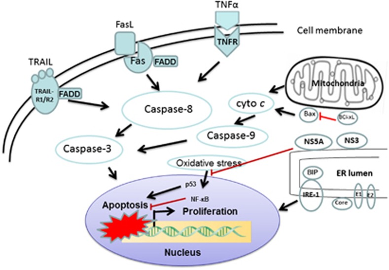
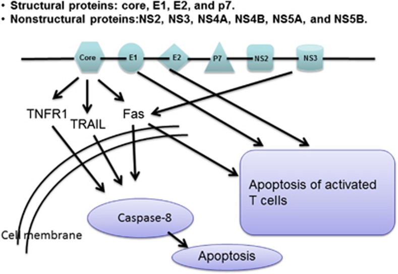
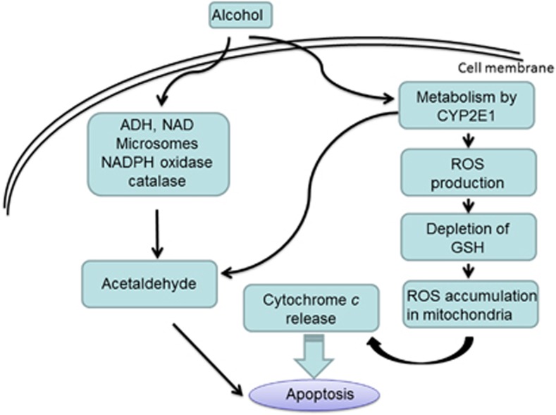
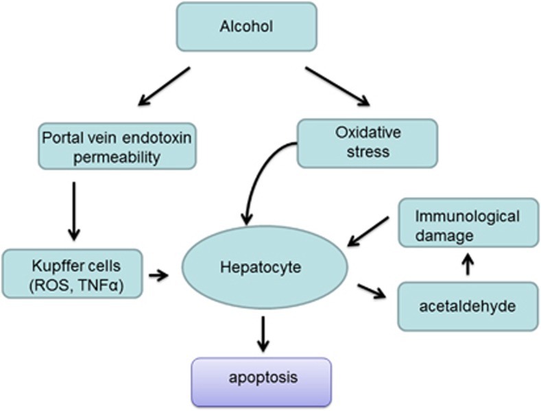
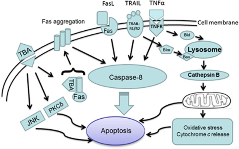
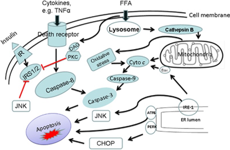
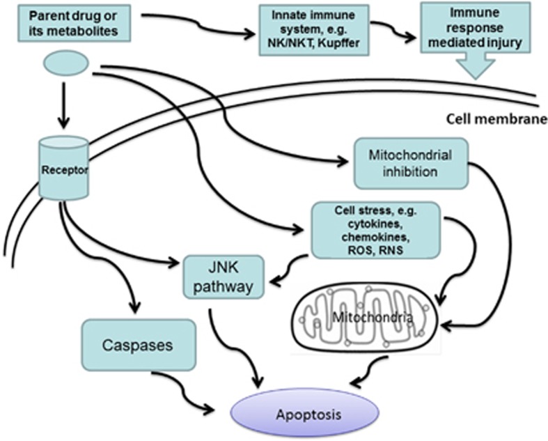
Apoptosis is a prominent feature of liver diseases. Causative factors such as alcohol, viruses, toxic bile acids, fatty acids, drugs, and immune response, can induce apoptotic cell death via membrane receptors and intracellular stress. Apoptotic signaling network, including membrane death receptor-mediated cascade, reactive oxygen species (ROS) generation, endoplasmic reticulum (ER) stress, lysosomal permeabilization, and mitochondrial dysfunction, is intermixed each other, but one mechanism may dominate at a particular stage. Mechanisms of hepatic apoptosis are complicated by multiple signaling pathways. The progression of liver disease is affected by the balance between apoptotic and antiapoptotic capabilities. Therapeutic options of liver injury are impacted by the clear understanding toward mechanisms of hepatic apoptosis.


Figures

Figure 1
Figure 1
Model of HCV apoptotic signaling pathways. HCV induces infected hepatocytes to apoptosis. Death receptor-mediated extrinsic pathway is enforced by mitochondrial amplification loop. Oxidative and ER stress with apoptosis are also shown, which reflect a potential interaction between the host cell response and apoptosis. Stress pathways are converged at the nucleus and low levels of NF-κB and BCL-xL sensitize hepatocytes to apoptosis. Lines with arrows denote an activating reaction; red lines ending in perpendicular lines denote inhibition of the reaction
Figure 2
Figure 2
Role of HCV viral proteins in the induction of apoptosis. The positive-stranded genome RNA of HCV encodes structural proteins (core, E1, E2, and p7) and non-structural proteins (NS2, NS3, NS4A, NS4B, NS5A, and NS5B). Special structures of HCV genome determine not only particular entry pattern of infectious HCV, but death mode of infected hepatocytes and immune escape mechanism as well
Figure 3
Figure 3
Molecular mechanism of alcohol-mediated hepatocyte apoptosis. Alcohol is metabolized in the liver and can be converted into acetaldehyde. As a toxic substance, the acetaldehyde induces hepatocyte apoptosis. Alcohol breakdown by cytochrome P450 2E1 (CYP2E1) also generates highly reactive molecules known as reactive oxygen species (ROS), particularly after chronic alcohol consumption. ROS can be eliminated by antioxidants such as glutathione (GSH). When cell's antioxidants are depleted and ROS accumulation reaches a critical threshold, mitochondrial damage occurs. This process leads to the release of cytochrome c from the mitochondria, which then activates caspases apoptotic pathway
Figure 4
Figure 4
Signaling mechanism of alcoholic liver injury. Oxidative stress is an important mechanism in alcoholic liver disease. Moreover, alcohol increases levels of bacterial endotoxin in the liver, which further activates liver Kupffer cells to produce inflammatory cytokines (e.g., TNFα) and/or chemokines (e.g., IL-8). The acetaldehyde interacts with proteins in the liver, forming compounds called adducts that can additionally activate certain immune cells to produce various cytokines, including interleukins, IFNγ, and TNFα. All of these diverse pathways contribute to the induction of apoptosis and organ damage
Figure 5
Figure 5
Toxic bile acids (TBAs)-induced hepatocyte apoptosis. In cholestasis, bile secretion is impaired, resulting in elevated concentrations of TBAs within hepatocytes. TBAs can activate the ER stress pathway as well as generation of ROS. TBAs also sensitize hepatocytes to death receptors (Fas, TRAIL, and TNFα)-induced apoptosis. Particularly, TBAs trigger translocation of intracellular Fas bearing vesicles to the plasma membrane where they self-aggregate, activate Fas receptor complexes, and cause caspase apoptotic cascade. In addition, some bile acids also activate PKC-δ and/or JNK1/2 MAP kinase, both of which stimulate pro-apoptotic pathways
Figure 6
Figure 6
Possible pathways of fat-induced hepatic apoptosis. The distinct mediators of fatty liver disease may include circulating cytokines, adipokines, and free fatty acids (FFAs). Peripheral insulin resistance enhances the delivery of FFAs to the liver, resulting in an imbalance of FFA metabolism and synthesis, thus promoting hepatic steatosis. Steatotic livers increase inflammatory cytokines, ROS, and ER stress, which trigger apoptotic cascade. Insulin sensitivity is further impaired by JNK-mediated phosphorylation of IRS1 and 2. Moreover, adipocytokines such as leptin and adiponectin are regulated by levels of circulating cytokine such as TNFα
Figure 7
Figure 7
Potential mechanisms for drug-induced hepatic apoptosis. Pathogenesis of drug-induced liver injury (DILI) includes cell stress, mitochondrial impairment, and specific immune reactions. Parent drugs or their reactive metabolites can cause cell stress to produce cytokines, chemokines, ROS, and reactive nitrogen species (RNS). Current concepts emphasize the central role of mitochondria, which leads to apoptotic and/or necrotic cell death. Hepatocytes, Kupffer cells, and even endothelial cells all participate in DILI. Inflammatory mediators of innate immune system determine the outcome of DILI
Figure 8
Figure 8
Pathogenesis of immune-mediated apoptotic liver injury. The liver has big population of Kupffer cells, dendritic cells, NK cells, and NKT cells. An innate immune response is initiated when antigens are presented by antigen-presenting cells (APCs) which then activate, directly and/or indirectly, NKT cells and other innate immune cells. The apoptosis of biliary epithelial cells (BECs) is a potential source of ‘neo-antigens' that may be responsible for facilitating molecular mimicry or autoimmunity. NKT cells upregulate FasL on their surface which binds to the Fas receptor expressed on target hepatocytes, leading to apoptosis. Activation of NKT cells can also indirectly induce hepatocyte apoptosis through the release of cytokines, including IFNγ and TNFα. Th2 responses are also induced due to the presence of IL-4, which then promote maturation of B cells into plasma cells for the production of autoantibodies. Furthermore, IL-17 family has been linked to many immune/autoimmune-related diseases

Similar articles

Cited by

References

    1. Smith PG, Tee LB, Yeoh GC. Appearance of oval cells in the liver of rats after long-term exposure to ethanol. Hepatology. 1996;23:145–154. - PubMed
    1. Sun C, Jin XL, Xiao JC. Oval cells in hepatitis B virus-positive and hepatitis C virus-positive liver cirrhosis: histological and ultrastructural study. Histopathology. 2006;48:546–555. - PubMed
    1. Soden JS, Devereaux MW, Haas JE, Gumpricht E, Dahl R, Gralla J, et al. Subcutaneous vitamin E ameliorates liver injury in an in vivo model of steatocholestasis. Hepatology. 2007;46:485–495. - PubMed
    1. Dahl TB, Haukeland JW, Yndestad A, Ranheim T, Gladhaug IP, Damas JK, et al. Intracellular nicotinamide phosphoribosyltransferase protects against hepatocyte apoptosis and is down-regulated in nonalcoholic fatty liver disease. J Clin Endocrinol Metab. 2010;95:3039–3047. - PubMed
    1. Waldhauser KM, Torok M, Ha HR, Thomet U, Konrad D, Brecht K, et al. Hepatocellular toxicity and pharmacological effect of amiodarone and amiodarone derivatives. J Pharmacol Exp Ther. 2006;319:1413–1423. - PubMed

Publication types

Substances