. 2014 Jan 16;5(1):e997.
doi: 10.1038/cddis.2013.533.

Calcineurin suppresses AMPK-dependent cytoprotective autophagy in cardiomyocytes under oxidative stress

Affiliations

Calcineurin suppresses AMPK-dependent cytoprotective autophagy in cardiomyocytes under oxidative stress

H He et al. Cell Death Dis. .

Abstract

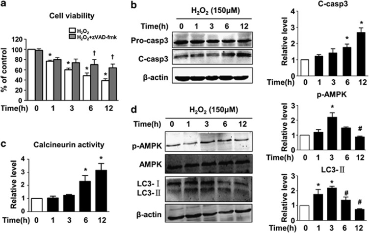
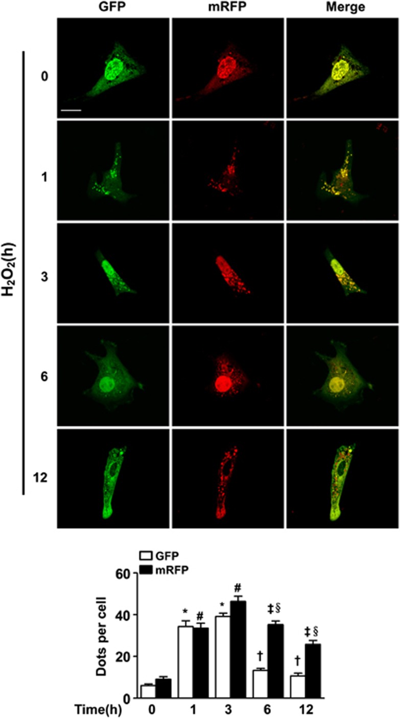
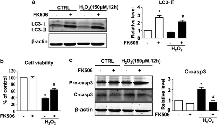
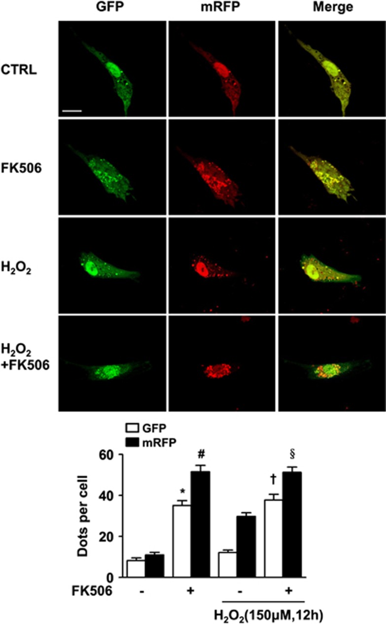
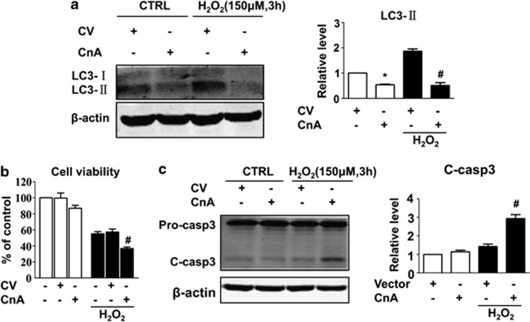
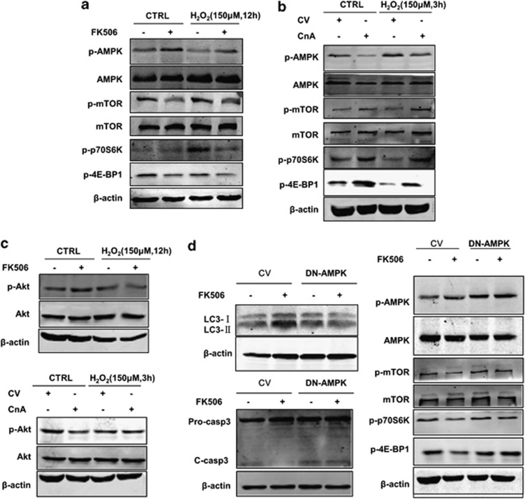
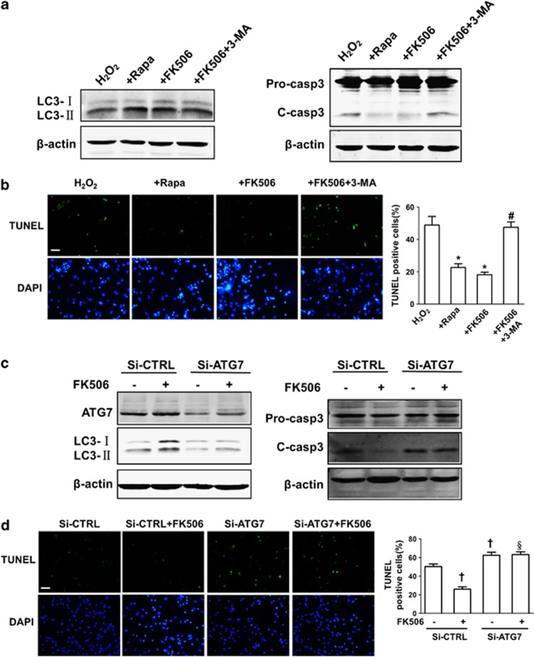
Calcineurin signalling plays a critical role in the pathogenesis of many cardiovascular diseases. Calcineurin has been proven to affect a series of signalling pathways and to exert a proapoptotic effect in cardiomyocytes. However, whether it is able to regulate autophagy remains largely unknown. Here, we report that prolonged oxidative stress-induced activation of calcineurin contributes to the attenuation of adaptive AMP-activated protein kinase (AMPK) signalling and inhibits autophagy in cardiomyocytes. Primary cardiomyocytes exhibited rapid formation of autophagosomes, microtubule-associated protein 1 light chain 3 (LC3) expression and phosphorylation of AMPK in response to hydrogen peroxide (H₂O₂) treatment. However, prolonged (12 h) H₂O₂ treatment attenuated these effects and was accompanied by a significant increase in calcineurin activity and apoptosis. Inhibition of calcineurin by FK506 restored AMPK function and LC3 expression, and decreased the extent of apoptosis caused by prolonged oxidative stress. In contrast, overexpression of the constitutively active form of calcineurin markedly attenuated the increase in LC3 induced by short-term (3 h) H₂O₂ treatment and sensitised cells to apoptosis. In addition, FK506 failed to induce autophagy and alleviate apoptosis in cardiomyocytes expressing a kinase-dead K45R AMPK mutant. Furthermore, inhibition of autophagy by 3-methylanine (3-MA) or by knockdown of the essential autophagy-related gene ATG7 abrogated the protective effect of FK506. These findings suggest a novel role of calcineurin in suppressing adaptive autophagy during oxidative stress by downregulating the AMPK signalling pathway. The results also provide insight into how altered calcineurin and autophagic signalling is integrated to control cell survival during oxidative stress and may guide strategies to prevent cardiac oxidative damage.


Figures

Figure 1
Figure 1
The effect of hydrogen peroxide (H2O2) on cardiomyocyte apoptosis, calcineurin activity, AMPK phosphorylation and LC3 expression. Primary cardiomyocytes were treated with H2O2 at a final concentration of 150 μM for 1, 3, 6 or 12 h. (a) The effect of H2O2 on cell viability at various time points with or without zVAD-fmk. (b) H2O2 induces time-dependent activation of caspase-3 as shown by western blotting. The statistical bar graph (right panel) shows the comparison of cleaved caspase-3 levels that have been normalised to β-actin. (c) H2O2 time dependently enhances calcineurin (Cn) activity. Cells were treated as described. Calcineurin activity in each group was normalised to the control group. (d) The effect of H2O2 on LC3 and AMPK protein expression at each time point as detected by western blotting. The levels of LC3-II and p-AMPK were normalised to β-actin and are shown in the bar graph (right panel). Data are presented as the mean±S.E.M., n≥3. *P<0.05 versus the control group, #P<0.05 versus 3 h and P<0.05 versus H2O2 alone at 6 and 12 h
Figure 2
Figure 2
Hydrogen peroxide (H2O2) promotes autophagic flux in cardiomyocytes. Cardiomyocytes were transfected with tandem-LC3 construct (GFP-mRFP-LC3) for 24 h, exposed to 150 μM H2O2 for 1, 3, 6 or 12 h and photographed using confocal microscopy. Representative images are shown in the upper panel. Both the GFP and mRFP dots were counted, and the statistical analyses are shown in the bottom panel. The data were obtained from 3 independent experiments, and 10 cells were scored in each experiment. Scale bar=20 μM. Data are presented as the mean±S.E.M. *P<0.05 versus control GFP, #P<0.05 versus control mRFP, P<0.05 versus 3 h GFP, P<0.05 versus 3 h mRFP, and §P<0.05 versus the corresponding GFP dots at each time point
Figure 3
Figure 3
FK506 induces autophagy and alleviates hydrogen peroxide (H2O2)-induced apoptosis. Cells were incubated with or without H2O2 (150 μM) in the presence or absence of FK506 (1 μM) for 12 h and subjected to MTT assay and western blotting, respectively. (a) FK506 induced LC3 expression with or without H2O2. LC3-II levels were normalised to β-actin and are shown in the bar graph on the right panel. (b) FK506 increased cell viability and (c) decreased the cleavage of caspase-3. The cleaved caspase-3 expression was normalised to β-actin and is shown in the right panel. Data are presented as the mean±S.E.M., n≥3. *P<0.05 versus the control group and #P<0.05 versus the H2O2 alone group
Figure 4
Figure 4
FK506 promotes the formation of autophagosomes and autolysosomes. Cells were transfected as described in Figure 2 and were subsequently treated as described in Figure 3. The number of both the GFP and mRFP dots was significantly increased with or without H2O2 treatment as shown in the bar graph in the bottom panel. Scale bar=20 μM. Data are presented as the mean±S.E.M. *P<0.05 versus control GFP, #P<0.05 versus control mRFP, P<0.05 versus H2O2-alone GFP and §P<0.05 versus H2O2-alone mRFP
Figure 5
Figure 5
Overexpression of calcineurin inhibits autophagy and sensitises cells to undergo H2O2-induced apoptosis. Cells were transfected with the control vector (CV) or vector expressing calcineurin A for 24 h and subsequently incubated with or without H2O2 (150 μM) for 3 h. After the treatment, the cells were harvested for MTT assay and western blotting. (a) Calcineurin inhibits LC3 expression whether H2O2 is present or not. (b) Overexpression of calcineurin results in decreased cell viability and (c) increased caspase-3 cleavage. Data are presented as the mean±S.E.M., n≥3. *P<0.05 versus the control group and #P<0.05 versus the H2O2-alone group
Figure 6
Figure 6
AMPK/mTOR signalling mediates the effect of calcineurin on autophagy and cell survival after H2O2 treatment. (a) Inhibition of calcineurin by FK506 enhances AMPK phosphorylation and attenuates the phosphorylation of mTOR. Cells were treated as described in Figure 3, and whole-cell lysates were subjected to western blot analysis. (b) Overexpression of calcineurin attenuates the phosphorylation of AMPK and enhances mTOR phosphorylation. Cells were treated as described in Figure 5, and whole-cell lysates were subjected to western blot analysis. (c) Calcineurin attenuates Akt signalling under normal conditions but not under oxidative stress conditions. Cells were treated as previously described. (d) FK506 showed no effect on autophagy regulation, cell protection and mTOR inhibition in DN-AMPK-expressing cells. Cells were transfected with the control vector (CV) and the DN-AMPK vector for 24 h and subsequently incubated with 150 μM H2O2 in the presence or absence of FK506 (1 μM) for 12 h. Representative images are from three or more independent experiments
Figure 7
Figure 7
Autophagy protects cells from apoptosis induced by hydrogen peroxide (H2O2). (a) The effect of rapamycin, FK506 and FK506 combined with 3-MA on LC3-II and cleaved caspase-3 expression as detected by western blotting. (b) The effect of rapamycin, FK506 and FK506 combined with 3-MA on H2O2-induced apoptosis as determined by TUNEL staining. Cells were incubated with H2O2 (150 μM) in the presence of rapamycin (Rapa, 500 nM), FK506 (1 μM) or FK506 (1 μM) combined with 3-MA (5 mM) for 12 h. (c) The effect of ATG7 knockdown on LC3-II and cleaved caspase-3 expression detected by western blotting. (d) The effect of ATG7 knockdown on H2O2-induced apoptosis as determined by TUNEL staining. Cells were transfected with negative control or ATG7 siRNA for 48 h and were subsequently subjected to H2O2 (150 μM) for 12 h with or without FK506 (1 μM). Scale bar=100 μM, the data for statistical analysis were from three independent experiments and the percentage of TUNEL-positive cells was scored in four non-overlapping microscopic fields in each experiment. Data are presented as the mean±S.E.M. *P<0.05 versus H2O2 alone and #P<0.05 versus H2O2+FK506, P<0.05 versus si-CTRL and §P<0.05 versus si-CTRL+FK506

References

    1. Dhalla NS, Temsah RM, Netticadan T. Role of oxidative stress in cardiovascular diseases. J Hypertens. 2000;18:655–673. - PubMed
    1. Ansley DM, Wang B. Oxidative stress and myocardial injury in the diabetic heart. J Pathol. 2013;229:232–241. - PMC - PubMed
    1. Kubli DA, Gustafsson AB. Mitochondria and mitophagy: the yin and yang of cell death control. Circ Res. 2012;111:1208–1221. - PMC - PubMed
    1. Kongara S, Karantza V. The interplay between autophagy and ROS in tumorigenesis. Front Oncol. 2012;2:171. - PMC - PubMed
    1. Levine B, Kroemer G. Autophagy in the pathogenesis of disease. Cell. 2008;132:27–42. - PMC - PubMed

Publication types