. 2014 Mar 15;127(Pt 6):1318-26.
doi: 10.1242/jcs.143545. Epub 2014 Jan 16.

A Pil1-Sle1-Syj1-Tax4 functional pathway links eisosomes with PI(4,5)P2 regulation

Affiliations

A Pil1-Sle1-Syj1-Tax4 functional pathway links eisosomes with PI(4,5)P2 regulation

Ruth Kabeche et al. J Cell Sci. .

Abstract

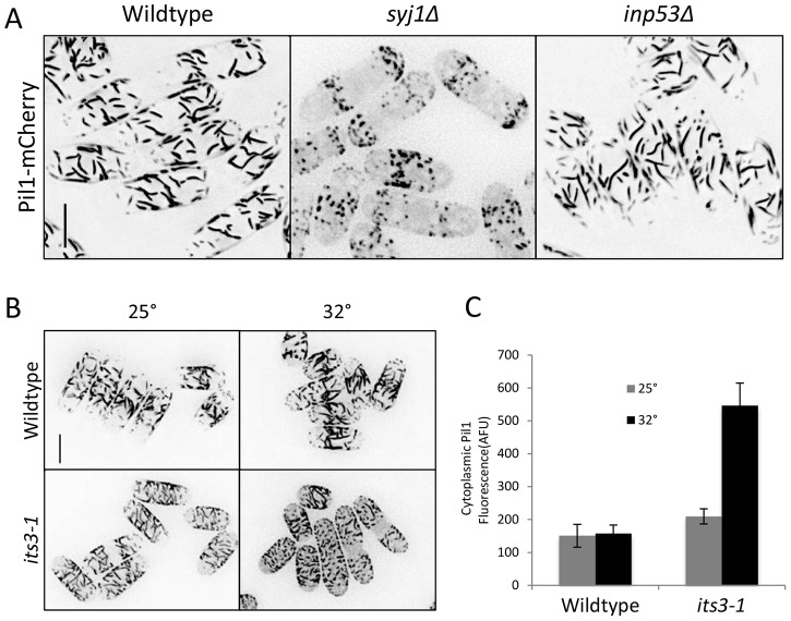
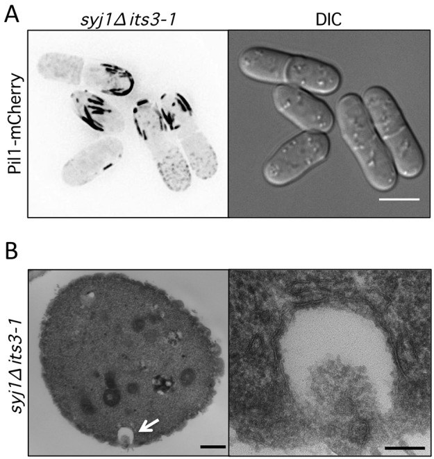
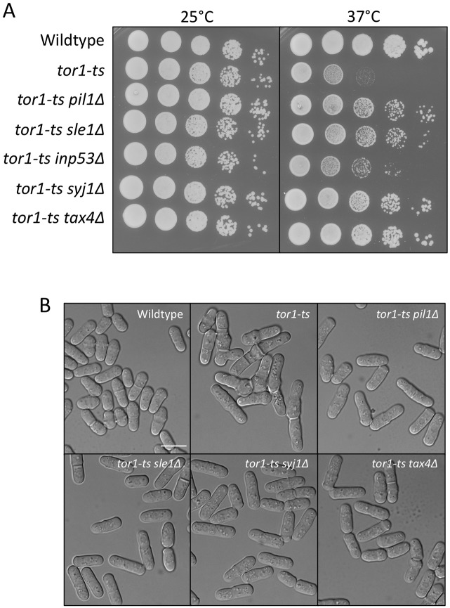
Stable compartments of the plasma membrane promote a wide range of cellular functions. In yeast cells, cytosolic structures called eisosomes generate prominent cortical invaginations of unknown function. Through a series of genetic screens in fission yeast, we found that the eisosome proteins Pil1 and Sle1 function with the synaptojanin-like lipid phosphatase Syj1 and its ligand Tax4. This genetic pathway connects eisosome function with the hydrolysis of phosphatidylinositol (4,5)-bisphosphate [PI(4,5)P2] in cells. Defects in PI(4,5)P2 regulation led to eisosome defects, and we found that the core eisosome protein Pil1 can bind to and tubulate liposomes containing PI(4,5)P2. Mutations in components of the Pil1-Sle1-Syj1-Tax4 pathway suppress the growth and morphology defects of TORC2 mutants, indicating that eisosome-dependent regulation of PI(4,5)P2 feeds into signal transduction pathways. We propose that the geometry of membrane invaginations generates spatial and temporal signals for lipid-mediated signaling events in cells.

Keywords: Eisosome; PI(4,5)P2; Synaptojanin; TORC2.


Figures

Fig. 1.
Fig. 1.
Genetic interactions identified by synthetic genetic array screens. (A) Summary of genetic interaction results for pil1Δ and sle1Δ; the table is limited to shared hits that were verified by tetrad dissection. (B) Description of key genes in Pil1-Sle1 genetic interaction screens. (C) Tetrad analysis confirms that pil1Δ, sle1Δ, syj1Δ, and tax4Δ are all synthetically lethal with inp53Δ. Inviable spores represented by dashed black squares; replica plating showed that inviable spores are the double mutants.
Fig. 2.
Fig. 2.
Eisosomes function in a genetic pathway for PI(4,5)P2 regulation. (A) Regulation of PI(4,5)P2 synthesis and hydrolysis in fission yeast. (B) Mutations in the Pil1–Sle1–Syj1–Tax4 pathway suppress temperature-sensitive growth defects of its3-1. (C) its3-1 suppresses the synthetic lethality of inp53Δ pil1Δ and inp53Δ syj1Δ mutants. Panels B and C are 10-fold serial dilutions grown at the indicated temperatures on rich media.
Fig. 3.
Fig. 3.
Eisosome defects in syj1 and its3 mutants. (A) Pil1 cortical filaments are dependent on Syj1 but not Inp53 for proper organization. Images are inverted maximum projections for Z-planes in the top half of the cell. Scale bar: 5 µm. (B) Eisosome organization is dependent on Its3. Cells were grown at 25°C and then switched to 32°C for 10 minutes. Images are inverted maximum projections from Z-planes in the top half of the cell. Scale bar, 5 µm. (C) Quantification of Pil1 cytoplasmic concentration in the indicated strain and temperature. Levels are presented as arbitrary fluorescence units (AFU) and represent mean ± s.d. for 25 cells.
Fig. 4.
Fig. 4.
Pil1 generates cortical pits in syj1Δ its3-1 double-mutant cells. (A) Thick Pil1 filaments at the cortex of syj1Δ its3-1 double-mutant cells. Images are inverted maximum projections from Z-planes in the top half of the cell. Scale bar, 5 µm. (B) Thin section electron microscopy of syj1Δ its3-1 double-mutant cells. Cross-section view displays exaggerated pit-like invagination. The white arrow highlights a pit-like invagination that is magnified in right panel. Scale bars: 500 nm (left panel); 100 nm (right panel).
Fig. 5.
Fig. 5.
Pil1 binds and tubulates liposomes in vitro. (A) Liposome pelleting assays. Pil1 was incubated with the indicated liposomes, and supernatant (S) and pellet (P) fractions were analyzed by SDS-PAGE followed by Coomassie staining. (B) Electron microscopy of negative-stained lipososomes containing PC/PS/PE/PI(4,5)2 in the presence or absence of purified Pil1. (C) Liposome pelleting assays. Pil1 was incubated with PC liposomes in the presence or absence of 1.5% PI(4,5)P2, and samples were analyzed as in panel A. (D) Electron microscopy of negative-stained samples from panel C.
Fig. 6.
Fig. 6.
Syj1 and Inp53 are spatially separated in cells. (A) Localization of endogenously tagged Syj1–mEGFP and Inp53–mEGFP. Images are inverted single focal planes from the cell middle. (B) Inp53–mEGFP does not colocalize with eisosomes, marked by Pil1–mCherry. (C) Most Syj1–mEGFP does not colocalize with eisosomes, marked by Pil1–mCherry. Arrows mark cortical puncta of Syj1–mEGFP. Images in B and C are inverted maximum projections from Z-planes in the top half of the cell, and also single focal planes from the cell middle. Scale bars: 5 µm.
Fig. 7.
Fig. 7.
Inp53 localizes to endocytic actin patches. (A) Colocalization of Inp53–mEGFP and Fim1–mCherry in actin patches; the region boxed in white is magnified in the bottom row. Images are inverted single focal planes. (B) Syj1–mEGFP does not colocalize with actin patches, marked by Fim1–mCherry; the region boxed in white is magnified in the bottom row. Images are inverted single focal planes. Scale bars, 5 µm.
Fig. 8.
Fig. 8.
Pil1–Sle1–Syj1–Tax4 pathway regulates TORC2 signaling. (A) 10-fold serial dilutions of the indicated tor1-ts single- and double-mutant cells at different temperatures. Note that temperature-sensitive growth of tor1-ts is suppressed by mutations in components of the Pil1–Sle1–Syj1–Tax4 pathway. (B) Digital interference contrast (DIC) images of tor1-ts single- and double-mutant cells. Cells were grown for 13 hours at 34°C. Scale bar, 10 µm.

Similar articles

Cited by

References

    1. Aguilar P. S., Fröhlich F., Rehman M., Shales M., Ulitsky I., Olivera-Couto A., Braberg H., Shamir R., Walter P., Mann M. et al. (2010). A plasma-membrane E-MAP reveals links of the eisosome with sphingolipid metabolism and endosomal trafficking. Nat. Struct. Mol. Biol. 17, 901–908 10.1038/nsmb.1829 - DOI - PMC - PubMed
    1. Bähler J., Wu J. Q., Longtine M. S., Shah N. G., McKenzie A., 3rd, Steever A. B., Wach A., Philippsen P., Pringle J. R. (1998). Heterologous modules for efficient and versatile PCR-based gene targeting in Schizosaccharomyces pombe. Yeast 14, 943–951 10.1002/(SICI)1097-0061(199807)14:10<943::AID-YEA292>3.0.CO;2-Y - DOI - PubMed
    1. Beltrao P., Cagney G., Krogan N. J. (2010). Quantitative genetic interactions reveal biological modularity. Cell 141, 739–745 10.1016/j.cell.2010.05.019 - DOI - PMC - PubMed
    1. Berchtold D., Piccolis M., Chiaruttini N., Riezman I., Riezman H., Roux A., Walther T. C., Loewith R. (2012). Plasma membrane stress induces relocalization of Slm proteins and activation of TORC2 to promote sphingolipid synthesis. Nat. Cell Biol. 14, 542–547 10.1038/ncb2480 - DOI - PubMed
    1. Brach T., Specht T., Kaksonen M. (2011). Reassessment of the role of plasma membrane domains in the regulation of vesicular traffic in yeast. J. Cell Sci. 124, 328–337 10.1242/jcs.078519 - DOI - PubMed

Publication types

MeSH terms

Substances