

JNK1 stress signaling is hyper-activated in high breast density and the tumor stroma: connecting fibrosis, inflammation, and stemness for cancer prevention
- PMID: 24434780
- PMCID: PMC3988118
- DOI: 10.4161/cc.27379
JNK1 stress signaling is hyper-activated in high breast density and the tumor stroma: connecting fibrosis, inflammation, and stemness for cancer prevention
Abstract
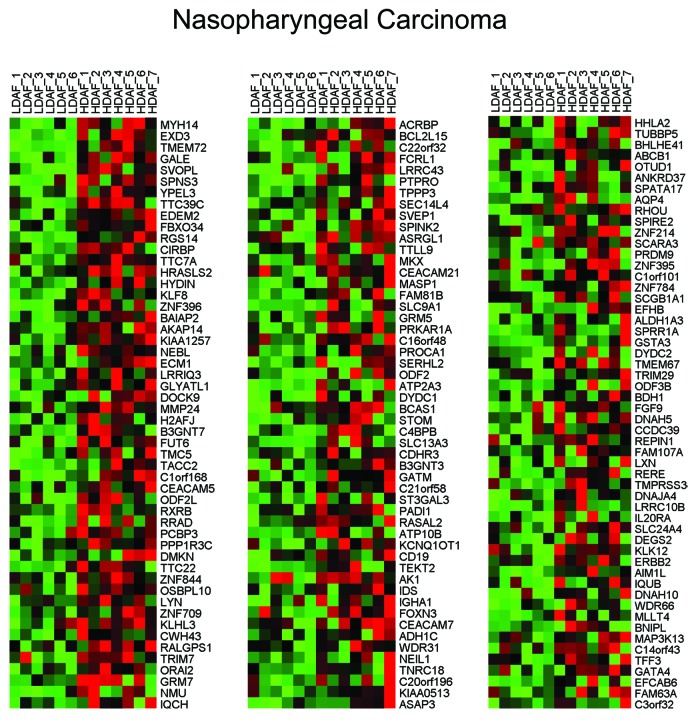
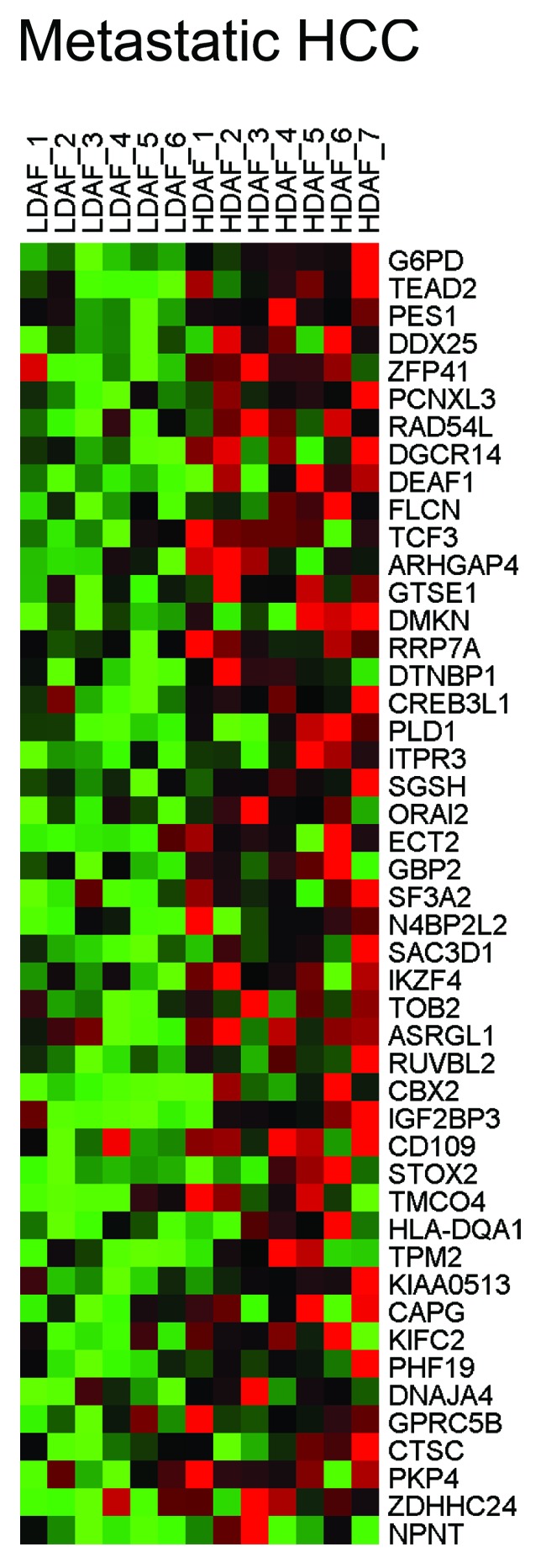
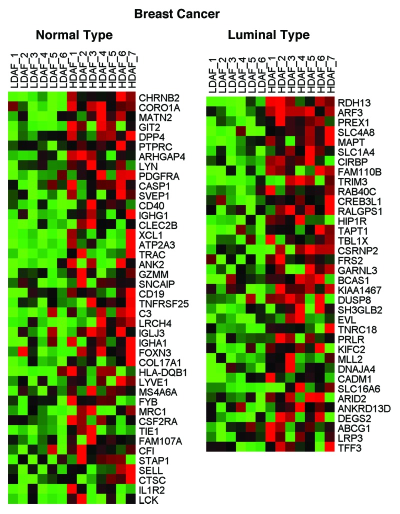
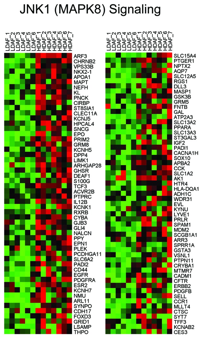
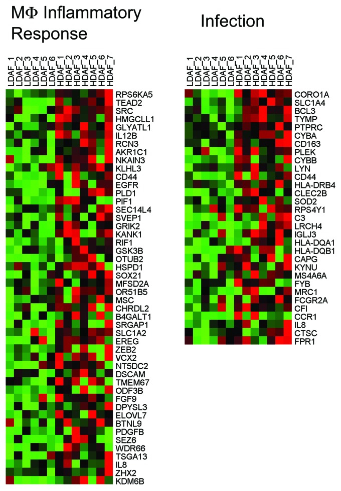
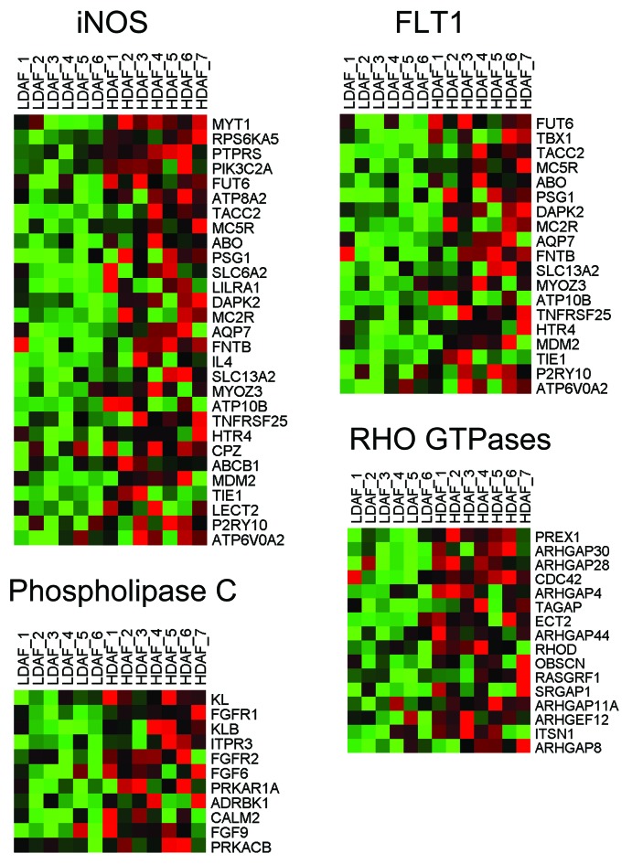
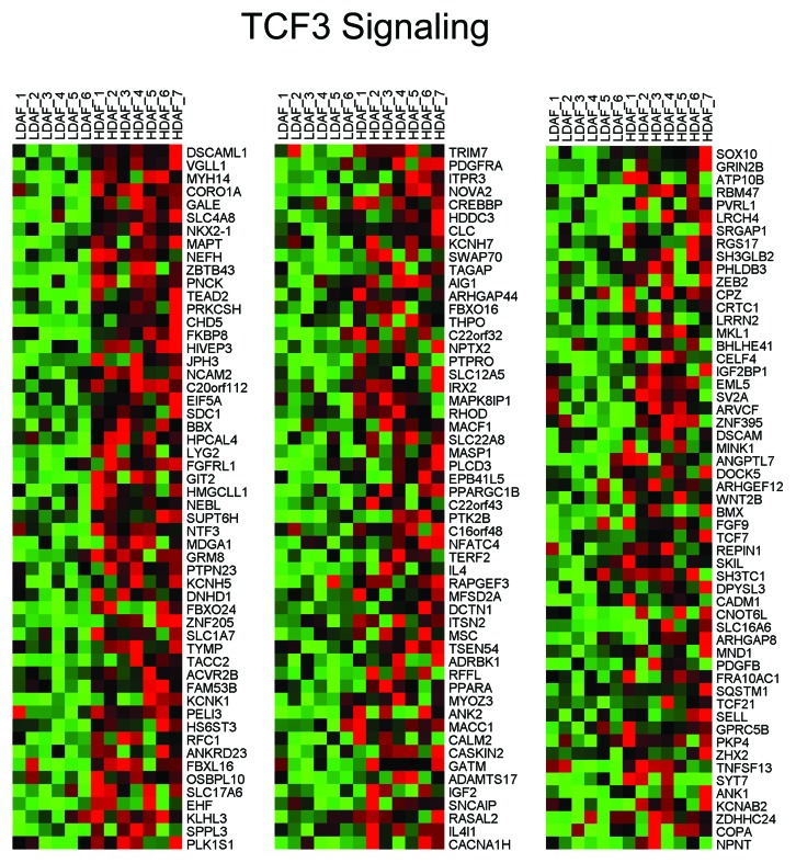
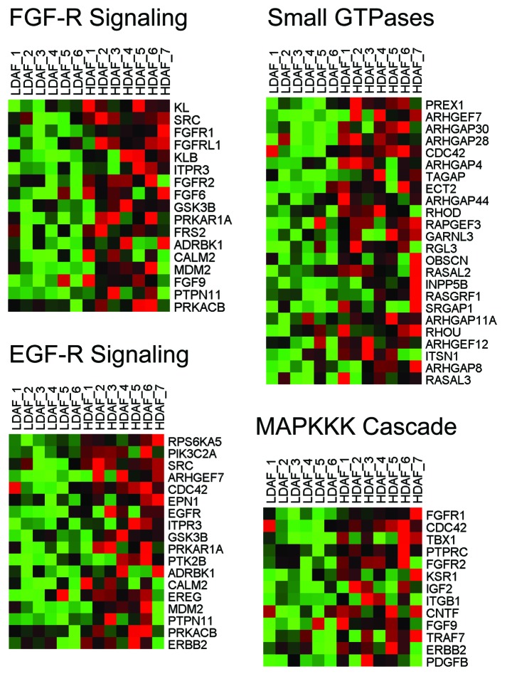
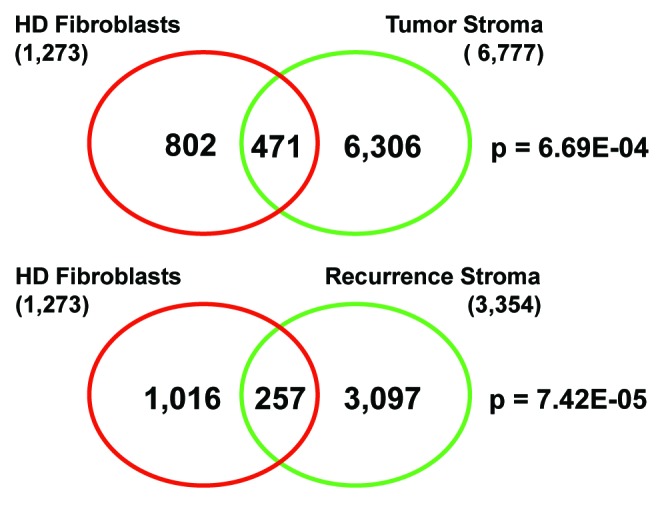
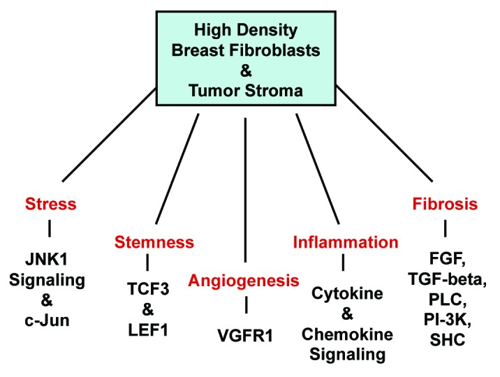
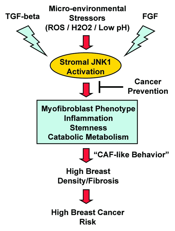
Mammography is an important screening modality for the early detection of DCIS and breast cancer lesions. More specifically, high mammographic density is associated with an increased risk of breast cancer. However, the biological processes underlying this phenomenon remain largely unknown. Here, we re-interrogated genome-wide transcriptional profiling data obtained from low-density (LD) mammary fibroblasts (n = 6 patients) and high-density (HD) mammary fibroblasts (n = 7 patients) derived from a series of 13 female patients. We used these raw data to generate a "breast density" gene signature consisting of>1250 transcripts that were significantly increased in HD fibroblasts, relative to LD fibroblasts. We then focused on the genes that were increased by ≥ 1.5-fold (P<0.05) and performed gene set enrichment analysis (GSEA), using the molecular signatures database (MSigDB). Our results indicate that HD fibroblasts show the upregulation and/or hyper-activation of several key cellular processes, including the stress response, inflammation, stemness, and signal transduction. The transcriptional profiles of HD fibroblasts also showed striking similarities to human tumors, including head and neck, liver, thyroid, lung, and breast cancers. This may reflect functional similarities between cancer-associated fibroblasts (CAFs) and HD fibroblasts. This is consistent with the idea that the presence of HD fibroblasts may be a hallmark of a pre-cancerous phenotype. In these biological processes, GSEA predicts that several key signaling pathways may be involved, including JNK1, iNOS, Rho GTPase(s), FGF-R, EGF-R, and PDGF-R-mediated signal transduction, thereby creating a pro-inflammatory, pro-proliferative, cytokine, and chemokine-rich microenvironment. HD fibroblasts also showed significant overlap with gene profiles derived from smooth muscle cells under stress (JNK1) and activated/infected macrophages (iNOS). Thus, HD fibroblasts may behave like activated myofibroblasts and macrophages, to create and maintain a fibrotic and inflammatory microenvironment. Finally, comparisons between the HD fibroblast gene signature and breast cancer tumor stroma revealed that JNK1 stress signaling is the single most significant biological process that is shared between these 2 data sets (with P values between 5.40E-09 and 1.02E-14), and is specifically associated with tumor recurrence. These results implicate "stromal JNK1 signaling" in the pathogenesis of human breast cancers and the transition to malignancy. Augmented TGF-β signaling also emerged as a common feature linking high breast density with tumor stroma and breast cancer recurrence (P = 5.23E-05). Similarities between the HD fibroblast gene signature, wound healing, and the cancer-associated fibroblast phenotype were also noted. Thus, this unbiased informatics analysis of high breast density provides a novel framework for additional experimental exploration and new hypothesis-driven breast cancer research, with a focus on cancer prevention and personalized medicine.
Keywords: EGF; FGF; JNK; PDGF; SAPK; TGF-beta; breast cancer; cancer associated fibroblasts; fibrosis; gene signature; inflammation; mammographic density; mammography; microenvironment; stress signaling; tumor stroma; wound healing.
Figures
Similar articles
-
Relationship of mammographic density and gene expression: analysis of normal breast tissue surrounding breast cancer.Clin Cancer Res. 2013 Sep 15;19(18):4972-4982. doi: 10.1158/1078-0432.CCR-13-0029. Epub 2013 Aug 5. Clin Cancer Res. 2013. PMID: 23918601 Free PMC article.
-
Gene expression in extratumoral microenvironment predicts clinical outcome in breast cancer patients.Breast Cancer Res. 2012 Mar 19;14(2):R51. doi: 10.1186/bcr3152. Breast Cancer Res. 2012. PMID: 22429463 Free PMC article.
-
Molecular profiling of a lethal tumor microenvironment, as defined by stromal caveolin-1 status in breast cancers.Cell Cycle. 2011 Jun 1;10(11):1794-809. doi: 10.4161/cc.10.11.15675. Epub 2011 Jun 1. Cell Cycle. 2011. PMID: 21521946 Free PMC article.
-
Regulation of heterogeneous cancer-associated fibroblasts: the molecular pathology of activated signaling pathways.J Exp Clin Cancer Res. 2020 Jun 16;39(1):112. doi: 10.1186/s13046-020-01611-0. J Exp Clin Cancer Res. 2020. PMID: 32546182 Free PMC article. Review.
-
Tumor-stromal interactions in breast tumor progression--significance of histological heterogeneity of tumor-stromal fibroblasts.Expert Opin Ther Targets. 2013 Apr;17(4):449-60. doi: 10.1517/14728222.2013.757305. Epub 2013 Jan 8. Expert Opin Ther Targets. 2013. PMID: 23297753 Review.
Cited by
-
JNK in Tumor Microenvironment: Present Findings and Challenges in Clinical Translation.Cancers (Basel). 2021 May 3;13(9):2196. doi: 10.3390/cancers13092196. Cancers (Basel). 2021. PMID: 34063627 Free PMC article. Review.
-
Key steps for effective breast cancer prevention.Nat Rev Cancer. 2020 Aug;20(8):417-436. doi: 10.1038/s41568-020-0266-x. Epub 2020 Jun 11. Nat Rev Cancer. 2020. PMID: 32528185 Review.
-
Role of immunosuppressive JNK pathway in the tumor microenvironment among TNBC subtypes in IBCSG trial 22-00.iScience. 2025 Jun 20;28(8):112964. doi: 10.1016/j.isci.2025.112964. eCollection 2025 Aug 15. iScience. 2025. PMID: 40822334 Free PMC article.
-
Stromal characteristics may hold the key to mammographic density: the evidence to date.Oncotarget. 2016 May 24;7(21):31550-62. doi: 10.18632/oncotarget.6912. Oncotarget. 2016. PMID: 26784251 Free PMC article. Review.
-
Use of Laplacian Heat Diffusion Algorithm to Infer Novel Genes With Functions Related to Uveitis.Front Genet. 2018 Oct 8;9:425. doi: 10.3389/fgene.2018.00425. eCollection 2018. Front Genet. 2018. PMID: 30349554 Free PMC article.
References
-
- Hattar R, Maller O, McDaniel S, Hansen KC, Hedman KJ, Lyons TR, Lucia S, Wilson RS, Jr., Schedin P. Tamoxifen induces pleiotrophic changes in mammary stroma resulting in extracellular matrix that suppresses transformed phenotypes. Breast Cancer Res. 2009;11:R5. doi: 10.1186/bcr2220. - DOI - PMC - PubMed
-
- Guo YP, Martin LJ, Hanna W, Banerjee D, Miller N, Fishell E, Khokha R, Boyd NF. Growth factors and stromal matrix proteins associated with mammographic densities. Cancer Epidemiol Biomarkers Prev. 2001;10:243–8. - PubMed
MeSH terms
Substances
Grants and funding
LinkOut - more resources
Full Text Sources
Other Literature Sources
Medical
Research Materials
Miscellaneous