

Surgical models for cardiac regeneration in neonatal mice
- PMID: 24434799
- PMCID: PMC3977725
- DOI: 10.1038/nprot.2014.021
Surgical models for cardiac regeneration in neonatal mice
Abstract
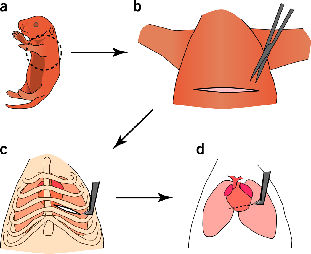
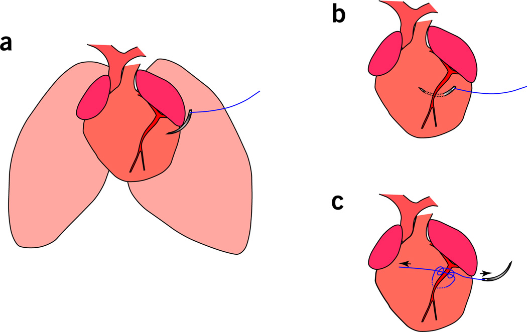
Although amphibian and fish models of heart regeneration have existed for decades, a mammalian equivalent has long remained elusive. Our discovery of a brief postnatal window for heart regeneration in neonatal mice has led to the establishment of surgical models for cardiac regenerative studies in mammals for the first time. This protocol describes a 10-min surgical procedure to induce cardiac injury in 1-d-old neonatal mice. This allows for the analysis of cardiac regeneration after surgical amputation of the left ventricle (LV) (apical resection) and coronary artery occlusion (myocardial infarction (MI)). A comparative analysis of neonatal and adult responses to myocardial injury should enable identification of the key differences between regenerative and nonregenerative responses to cardiac injury. This protocol can also be adapted to the growing repertoire of genetic models available in the mouse, and it provides a valuable tool for unlocking the molecular mechanisms that guide mammalian heart regeneration during early postnatal life.
Figures
References
-
- Jessup M, Brozena S. Heart failure. New Engl. J. Med. 2003;348:2007–2018. - PubMed
-
- Sutton MG, Sharpe N. Left ventricular remodeling after myocardial infarction: pathophysiology and therapy. Circulation. 2000;101:2981–2988. - PubMed
-
- Murry CE, Reinecke H, Pabon LM. Regeneration gaps: observations on stem cells and cardiac repair. J. Am. Coll. Cardiol. 2006;47:1777–1785. - PubMed
Publication types
MeSH terms
Grants and funding
LinkOut - more resources
Full Text Sources
Other Literature Sources
Medical
