

Comparison between PET template-based method and MRI-based method for cortical quantification of florbetapir (AV-45) uptake in vivo
- PMID: 24435769
- PMCID: PMC3978219
- DOI: 10.1007/s00259-013-2656-8
Comparison between PET template-based method and MRI-based method for cortical quantification of florbetapir (AV-45) uptake in vivo
Abstract
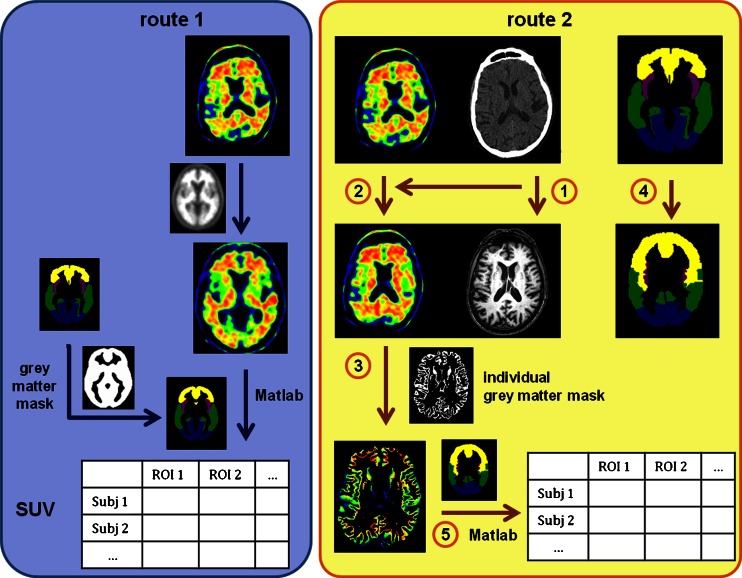
Purpose: Florbetapir (AV-45) has been shown to be a reliable tool for assessing in vivo amyloid load in patients with Alzheimer's disease from the early stages. However, nonspecific white matter binding has been reported in healthy subjects as well as in patients with Alzheimer's disease. To avoid this issue, cortical quantification might increase the reliability of AV-45 PET analyses. In this study, we compared two quantification methods for AV-45 binding, a classical method relying on PET template registration (route 1), and a MRI-based method (route 2) for cortical quantification.
Methods: We recruited 22 patients at the prodromal stage of Alzheimer's disease and 17 matched controls. AV-45 binding was assessed using both methods, and target-to-cerebellum mean global standard uptake values (SUVr) were obtained for each of them, together with SUVr in specific regions of interest. Quantification using the two routes was compared between the clinical groups (intragroup comparison), and between groups for each route (intergroup comparison). Discriminant analysis was performed.
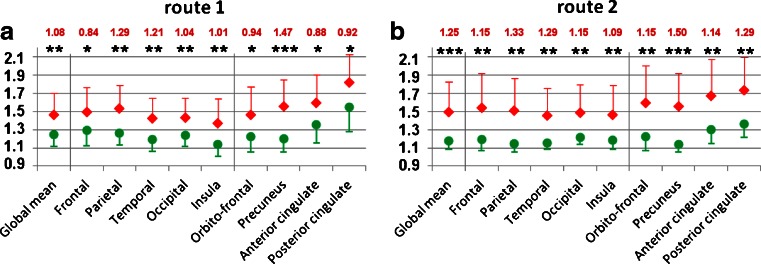
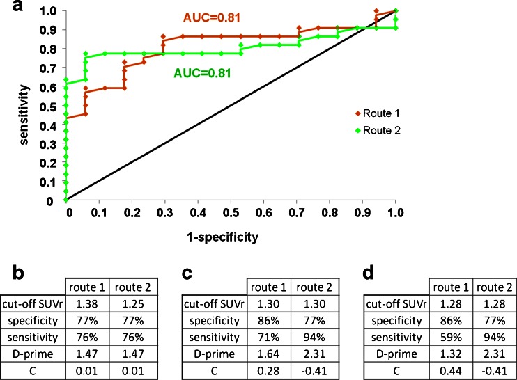
Results: In the intragroup comparison, differences in uptake values were observed between route 1 and route 2 in both groups. In the intergroup comparison, AV-45 uptake was higher in patients than controls in all regions of interest using both methods, but the effect size of this difference was larger using route 2. In the discriminant analysis, route 2 showed a higher specificity (94.1 % versus 70.6 %), despite a lower sensitivity (77.3 % versus 86.4 %), and D-prime values were higher for route 2.
Conclusion: These findings suggest that, although both quantification methods enabled patients at early stages of Alzheimer's disease to be well discriminated from controls, PET template-based quantification seems adequate for clinical use, while the MRI-based cortical quantification method led to greater intergroup differences and may be more suitable for use in current clinical research.
Figures
Similar articles
-
Perfusion-like template and standardized normalization-based brain image analysis using 18F-florbetapir (AV-45/Amyvid) PET.Eur J Nucl Med Mol Imaging. 2013 Jun;40(6):908-20. doi: 10.1007/s00259-013-2350-x. Epub 2013 Feb 15. Eur J Nucl Med Mol Imaging. 2013. PMID: 23412134
-
A Semiautomated Method for Quantification of F 18 Florbetapir PET Images.J Nucl Med. 2015 Nov;56(11):1736-41. doi: 10.2967/jnumed.114.153494. Epub 2015 Sep 3. J Nucl Med. 2015. PMID: 26338898
-
Comparison of MRI based and PET template based approaches in the quantitative analysis of amyloid imaging with PIB-PET.Neuroimage. 2013 Apr 15;70:423-33. doi: 10.1016/j.neuroimage.2012.12.014. Epub 2012 Dec 20. Neuroimage. 2013. PMID: 23261639
-
Quantification of 18F-florbetapir PET: comparison of two analysis methods.Eur J Nucl Med Mol Imaging. 2015 Apr;42(5):725-32. doi: 10.1007/s00259-015-2988-7. Epub 2015 Feb 5. Eur J Nucl Med Mol Imaging. 2015. PMID: 25652817
-
Positron emission tomography radiopharmaceuticals for imaging brain Beta-amyloid.Semin Nucl Med. 2011 Jul;41(4):283-99. doi: 10.1053/j.semnuclmed.2011.02.005. Semin Nucl Med. 2011. PMID: 21624562 Review.
Cited by
-
Low-dose CT for the spatial normalization of PET images: A validation procedure for amyloid-PET semi-quantification.Neuroimage Clin. 2018 Jul 19;20:153-160. doi: 10.1016/j.nicl.2018.07.013. eCollection 2018. Neuroimage Clin. 2018. PMID: 30094164 Free PMC article.
-
Amyloid PET quantification using low-dose CT-guided anatomic standardization.EJNMMI Res. 2021 Dec 14;11(1):125. doi: 10.1186/s13550-021-00867-7. EJNMMI Res. 2021. PMID: 34905145 Free PMC article.
-
Evaluation of software tools for automated identification of neuroanatomical structures in quantitative β-amyloid PET imaging to diagnose Alzheimer's disease.Eur J Nucl Med Mol Imaging. 2016 Jun;43(6):1077-87. doi: 10.1007/s00259-015-3300-6. Epub 2016 Jan 7. Eur J Nucl Med Mol Imaging. 2016. PMID: 26739328
-
Cognitive and functional patterns of nondemented subjects with equivocal visual amyloid PET findings.Eur J Nucl Med Mol Imaging. 2015 Aug;42(9):1459-68. doi: 10.1007/s00259-015-3067-9. Epub 2015 May 8. Eur J Nucl Med Mol Imaging. 2015. PMID: 25952279 Clinical Trial.
-
Associations between Aβ deposition and neurometabolic alterations in Alzheimer's disease: Insights from hybrid 3D MRSI-PET imaging.Alzheimers Dement. 2025 Jun;21(6):e70332. doi: 10.1002/alz.70332. Alzheimers Dement. 2025. PMID: 40495577 Free PMC article.
References
-
- McKhann G, Drachman D, Folstein M, Katzman R, Price D, Stadlan EM. Clinical diagnosis of Alzheimer’s disease: report of the NINCDS-ADRDA Work Group under the auspices of Department of Health and Human Services Task Force on Alzheimer’s Disease. Neurology. 1984;34:939–944. doi: 10.1212/WNL.34.7.939. - DOI - PubMed
-
- McKhann GM, Knopman DS, Chertkow H, Hyman BT, Jack CR, Jr, Kawas CH, et al. The diagnosis of dementia due to Alzheimer’s disease: recommendations from the National Institute on Aging–Alzheimer’s Association workgroups on diagnostic guidelines for Alzheimer’s disease. Alzheimers Dement. 2011;7:263–269. doi: 10.1016/j.jalz.2011.03.005. - DOI - PMC - PubMed
Publication types
MeSH terms
Substances
LinkOut - more resources
Full Text Sources
Other Literature Sources
Medical
