. 2014 Mar;4(3):334-47.
doi: 10.1158/2159-8290.CD-13-0611. Epub 2014 Jan 16.

Rapid induction of apoptosis by PI3K inhibitors is dependent upon their transient inhibition of RAS-ERK signaling

Affiliations

Rapid induction of apoptosis by PI3K inhibitors is dependent upon their transient inhibition of RAS-ERK signaling

Marie Will et al. Cancer Discov. 2014 Mar.

Abstract

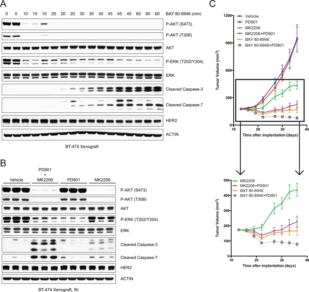
The effects of selective phosphoinositide 3-kinase (PI3K) and AKT inhibitors were compared in human tumor cell lines in which the pathway is dysregulated. Both caused inhibition of AKT, relief of feedback inhibition of receptor tyrosine kinases, and growth arrest. However, only the PI3K inhibitors caused rapid induction of cell death. In seeking a mechanism for this phenomenon, we found that PI3K inhibition, but not AKT inhibition, causes rapid inhibition of wild-type RAS and of RAF-MEK-ERK signaling. Inhibition of RAS-ERK signaling is transient, rebounding a few hours after drug addition, and is required for rapid induction of apoptosis. Combined MEK and AKT inhibition also promotes cell death, and in murine models of HER2(+) cancer, either pulsatile PI3K inhibition or combined MEK and AKT inhibition causes tumor regression. We conclude that PI3K is upstream of RAS and AKT and that pulsatile inhibition of both pathways is sufficient for effective antitumor activity.


Conflict of interest statement

CONFLICT OF INTEREST

Claudia Schneider and Ningshu Liu are employees of Bayer Research Labs

No other author has a conflict of interest to disclose.

Figures

Figure 1
Figure 1. PI3K inhibition induces more apoptosis than AKT inhibition
(A) BT-474 cells were treated with BAY 80-6946 (50nM) or MK2206 (2μM) and collected at indicated times. Immunoblots of AKT, AKT substrates, and surrogates for apoptosis show similar AKT inhibition but greater apoptosis with PI3K inhibition. (B) Cell cycle distribution of BT-474 cells treated with DMSO, MK2206 (2μM), or BAY 80-6946 (50nM) for 24, 48, or 72 hours as determined by FACS analysis show drugs causing similar suppression of S phase distribution. Apoptosis results were reported as mean percent SubG1 fraction with standard errors and show greater %SubG1 with PI3K inhibition. (C) BT-474 cells were treated with DMSO, MK2206 (2μM), or BAY 80-6946 (50nM) and viable cells were counted at indicated times, demonstrating loss in viable cell number with PI3K inhibition. Results were reported as a mean of triplicate with standard errors. (D) BT-474 cells untransfected or stably transfected with either pQCXIP vector or pQCXIP-BCL-XL were treated with BAY 80-6946 (50nM) and collected at indicated times. Immunoblots show BCL-XL overexpression abolishes the induction of cleaved caspase-3, cleaved-caspase-7 and cleaved PARP. (E) BT-474 cells stably transfected with pQCXIP vector or pQCXIP-BCL-XL and treated with DMSO or BAY 80-6946 (50nM) show that the decrease in viable cells by BAY 80-6946 is blocked by BCL-XL overexpression.
Figure 2
Figure 2. PI3K, but not AKT or mTOR, is upstream of the ERK signaling pathway
(A) BT-474 cells were treated with BAY 80-6946 (50nM) or MK2206 (2μM) and collected at indicated times. Immunoblots of AKT and mTOR substrates, RTKs, and parallel pathways like STAT and ERK. (B, C, D) BT-474 cells were treated with BAY 80-6946 (50nM) (B), or MK2206 (2μM) (C), or AZD8055 (250nM) (D), and collected at indicated times. Immunoblots show downregulation of P-ERK by PI3K, but not AKT or mTOR, inhibition.
Figure 3
Figure 3. PI3K isoform selective inhibitors and PTEN induction decrease ERK phosphorylation
(A) BT-474 cells were treated with the PI3Kα specific inhibitor BYL-719 (5μM) (left); MDA-MB-468 cells were treated with the PI3Kβ specific inhibitor GSK2311418A (1μM) (center); CCRF-SB cells were treated with the PI3Kδ specific inhibitor CAL101 (1μM) (right). (B) PTEN expression was induced in MDA-MB-468TR-PTEN cells by doxycycline (100ng/mL). (A, B) Cells were collected at indicated times. Immunoblots show loss of phosphorylation of RAF/MEK/ERK with inhibition of PI3K isoforms or induction of PTEN. N.S. denotes non-specific band.
Figure 4
Figure 4. PI3K regulates ERK signaling in many tumor models
Cell lines with indicated tumor genotype were treated with BAY 80-6946 (50nM) and collected at indicated times. Immunoblots show downregulation of P-AKT in all cell lines and loss of P-ERK in the majority of RAS wild type cells.
Figure 5
Figure 5. PI3K regulates the level of active RAS in RAS wild type cells
(A) SK-BR-3 cells (left) or BT-474 cells (right) were treated with BAY 80-6946 (50nM) and collected at indicated times. RAS-GTP level was assessed by RAF1-RBD pull down (PD) and lysates from the same samples were immunoblotted. A control for the PD (Neg. Ctrl.) was performed in the same manner as the other samples but without the addition of RAF1-RBD. Immunoblots demonstrate that PI3K inhibition blocks AKT phosphorylation and causes a loss of RAS-GTP level (total and individual isoforms) corresponding with changes in RAF-MEK-ERK activity. (B) SK-BR-3 cells (left) or BT-474 cells (right) were treated with BAY 80-6946 (50nM) or MK2206 (2μM) for 30 minutes, EGF (100ng/mL) for 5 minutes, Lapatinib (2μM) for 1h, or left untreated, and collected for RAS-GTP analysis (as in (A)) or immunoblotting. Treatment with EGF activates RAS and RAF/MEK/ERK, while RAS-ERK signaling is inhibited by RTK or PI3K inhibition but not AKT inhibition. (C) SK-BR-3 cells stably transfected with TTIGp-MLUEX-NRAS Q61K were pretreated for 24h with doxycycline (1μg/mL) to induce the expression of exogenous NRAS Q61K (right) or not pretreated (left) before the addition of the PI3K inhibitor BAY 80-6946 (50nM) for indicated times, EGF (100ng/mL) for 5 minutes, or Lapatinib (2μM) for 1h. RAS-GTP analysis was performed as in (A), and lysates were immunoblotted. PI3K inhibition decreases the GTP-bound level of the endogenous wild type, but not the exogenous mutant, RAS protein. Accordingly, P-ERK was only decreased in cells without the induction of NRAS Q61K expression. N.S. denotes non-specific band.
Figure 6
Figure 6. Rapid induction of apoptosis by PI3K inhibitor is dependent on its downregulation of P-ERK
(A) BT-474 cells were left untreated (lane 1), treated with BAY 80-6946 (50nM) alone (lanes 2, 3), or BAY 80-6946 (50nM) and EGF (100ng/mL) (lanes 4, 5), or BAY 80-6946 (50nM), EGF (100ng/mL) and PD901 (50nM) (lanes 6, 7), or PD901 (50nM) alone (lanes 8, 9), and collected at indicated times. Immunoblots showing greatest induction of cleaved caspase-3, cleaved caspase-7 and cleaved-PARP when both P-ERK and P-AKT are inhibited. (B) BT-474 cells were treated with MK2206 (2μM) alone, PD901 (50nM) alone, or a combination of MK2206 (2μM) and PD901 (50nM), and collected at indicated times. Immunoblots showing greatest induction of cleaved caspase-3 and cleaved caspase-7 by the combination of AKT and MEK inhibition. (C) BT-474 cells (top), SK-BR-3 cells (middle) or MDA-MB-361 cells (bottom) were treated in triplicate with DMSO, MK2206 (2μM), PD901 (50nM), BAY 80-6946 (50nM), a combination of MK2206 (2μM) and PD901 (50nM), or a combination of BAY 80-6946 (50nM) and PD901 (50nM), and collected at 72h. Samples were analyzed for induction of apoptosis by FACS, and results were reported as mean percent SubG1 fraction with standard errors (n=3/group).
Figure 7
Figure 7. Combined MEK and AKT inhibition improves antitumor efficacy in vivo
(A) Mice bearing BT-474 xenograft were treated with a single dose of BAY 80-6946 (14mg/kg) and tumors were collected at indicated times post-treatment. Immunoblots of tumor lysates demonstrate downregulation of P-AKT and P-ERK, as well as induction of cleaved caspase-3 and cleaved caspase-7. (B) Mice bearing BT-474 xenograft were treated with vehicle, or a combination of MK2206 (100mg/kg and PD901 (5mg/kg), or either agent alone, and tumors were collected at 5h post-treatment. Immunoblots of tumor lysates demonstrate greatest induction of cleaved caspase-3 and cleaved caspase-7 with the combination of MK2206 and PD901. (C) Mice bearing BT-474 xenograft were randomized to (1) Vehicle five times/week, (2) PD901 5mg/kg five times/week, (3) MK2206 100mg/kg five times/week, (4) BAY 80-6946 14mg/kg three times/week, (5) combination of (2) and (3), or (6) combination of (2) and (4), and tumor size was measured by vernier caliper two times/week. The results were reported as mean tumor volume with standard errors (n=10/group). Inset excludes group (1) and (2).

Comment in

References

    1. Cully M, You H, Levine AJ, Mak TW. Beyond PTEN mutations: the PI3K pathway as an integrator of multiple inputs during tumorigenesis. Nat Rev Cancer. 2006;6:184–192. - PubMed
    1. Saal LH, Holm K, Maurer M, Memeo L, Su T, Wang X, et al. PIK3CA mutations correlate with hormone receptors, node metastasis, and ERBB2, and are mutually exclusive with PTEN loss in human breast carcinoma. Cancer research. 2005;65:2554–2559. - PubMed
    1. Samuels Y, Wang Z, Bardelli A, Silliman N, Ptak J, Szabo S, et al. High frequency of mutations of the PIK3CA gene in human cancers. Science. 2004;304:554. - PubMed
    1. Vivanco I, Sawyers CL. The phosphatidylinositol 3-Kinase AKT pathway in human cancer. Nat Rev Cancer. 2002;2:489–501. - PubMed
    1. The Cancer Genome Atlas N, Genome sequencing centres: Washington University in St L. Koboldt DC, Fulton RS, McLellan MD, Schmidt H, et al. Comprehensive molecular portraits of human breast tumours. Nature. 2012 - PMC - PubMed

Publication types

MeSH terms

LinkOut - more resources