. 2014;29(5):1131-40.
doi: 10.1002/jbmr.2143.

Cortical and trabecular bone benefits of mechanical loading are maintained long term in mice independent of ovariectomy

Affiliations

Cortical and trabecular bone benefits of mechanical loading are maintained long term in mice independent of ovariectomy

Stuart J Warden et al. J Bone Miner Res. 2014.

Abstract

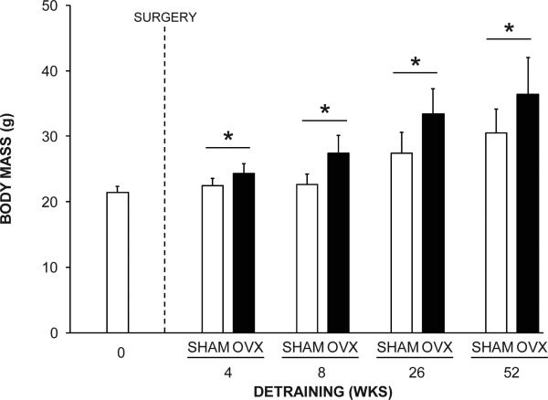
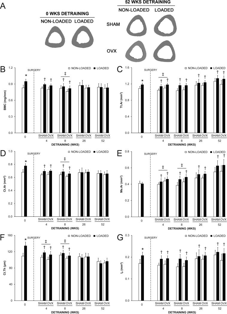
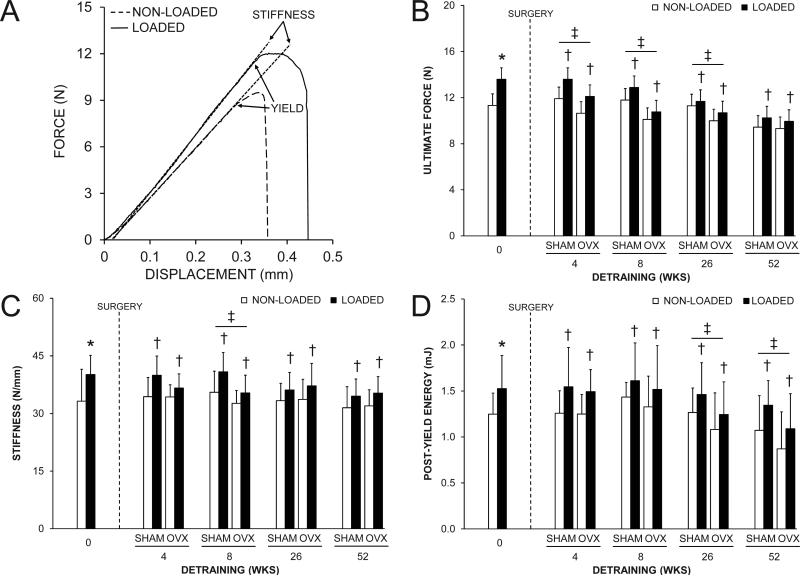
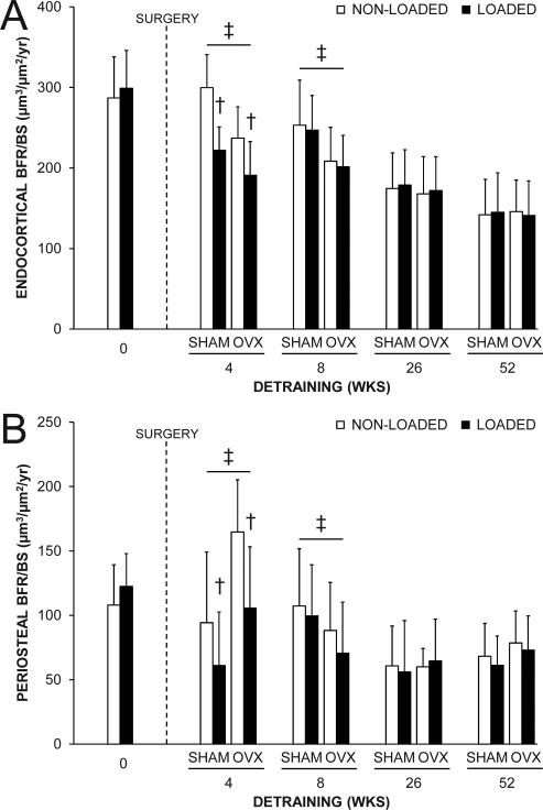
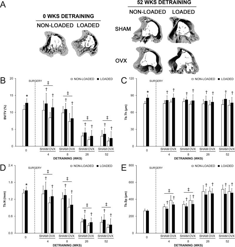
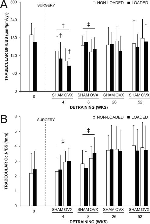
Skeletal loading enhances cortical and trabecular bone properties. How long these benefits last after loading cessation remains an unresolved, clinically relevant question. This study investigated long-term maintenance of loading-induced cortical and trabecular bone benefits in female C57BL/6 mice and the influence of a surgically induced menopause on the maintenance. Sixteen-week-old animals had their right tibia extrinsically loaded 3 days/week for 4 weeks using the mouse tibial axial compression loading model. Left tibias were not loaded and served as internal controls. Animals were subsequently detrained (restricted to cage activities) for 0, 4, 8, 26, or 52 weeks, with ovariectomy (OVX) or sham-OVX surgery being performed at 0 weeks detraining. Loading increased midshaft tibia cortical bone mass, size, and strength, and proximal tibia bone volume fraction. The cortical bone mass, area, and thickness benefits of loading were lost by 26 weeks of detraining because of heightened medullary expansion. However, loading-induced benefits on bone total area and strength were maintained at each detraining time point. Similarly, the benefits of loading on bone volume fraction persisted at all detraining time points. The long-term benefits of loading on both cortical and trabecular bone were not influenced by a surgically induced menopause because there were no interactions between loading and surgery. However, OVX had independent effects on cortical bone properties at early (4 and 8 weeks) detraining time points and trabecular bone properties at all detraining time points. These cumulative data indicate loading has long-term benefits on cortical bone size and strength (but not mass) and trabecular bone morphology, which are not influenced by a surgically induced menopause. This suggests skeletal loading associated with physical activity may provide long-term benefits by preparing the skeleton to offset both the cortical and trabecular bone changes associated with aging and menopause.

Keywords: EXERCISE; GROWTH AND DEVELOPMENT; MENOPAUSE; OSTEOPOROSIS; PHYSICAL ACTIVITY.


Figures

Fig. 1
Fig. 1
The effect of surgical intervention on body mass. OVX animals had greater body mass than SHAM animals in the 4, 8, 26 and 52 wks detraining groups (*p < 0.001). Data represent mean ± SD.
Fig. 2
Fig. 2
The effect of loading and surgery at select detraining time points on the midshaft tibia. A) Representative micro-CT tomographic images of the midshaft tibia in non-loaded and loaded bones from the 0 and 52 wks detraining groups. Loading increased total (Tt.Ar) and cortical (Ct.Ar) areas, and cortical thickness (Ct.Th), as evident in the 0 wks detraining group. The loading-induced increase in Tt.Ar persisted in the 52 wks detraining group in both SHAM and OVX animals. B) Bone mineral content (BMC); C) Tt.Ar; D) Ct.Ar; E) medullary area (Me.Ar); F) Ct.Th and G) polar moment of inertia (IP) at the midshaft tibia as select detraining time points. Loading increased BMC, Tt.Ar, Ct.Ar, Ct.Th and IP, as assessed in the 0 wks detraining group (*p < 0.05). There were no statistical interactions between loading and surgery in any detraining time point group. Loaded tibias had more BMC, Ct.Ar and Ct.Th in the 4 and 8 wks detraining groups and more Tt.Ar, Me.Ar and IP in each detraining time point group than non-loaded tibias (p < 0.05 for loading main effect). OVX animals had more Tt.Ar and Me.Ar, and less Ct.Th than SHAM animals in the 4 wks detraining group, and less BMC, Ct.Ar and Ct.Th, and more Me.Ar than SHAM animals in the 8 wks detraining group, (p < 0.05 for surgery main effect). Data represent body mass corrected means ± SD.
Fig. 3
Fig. 3
The effect of loading and surgery at select detraining time points on midshaft tibia mechanical properties. A) Representative force vs. displacement curves for a pair of loaded and non-loaded tibias from the 0 wks detraining group. Loading increased: A) ultimate force (peak of the curve on the y-axis in panel A); C) stiffness (slope of the linear portion of the curve in panel A), and; D) post-yield energy to failure (area under curve between yield point and failure in panel A), as assessed in the 0 wks detraining group (*p < 0.05). There were no statistical interactions between loading and surgery in any detraining time point group for any of the properties assessed. Loaded tibias had greater ultimate force, stiffness and post-yield energy to failure than non-loaded tibias in each detraining time point group (p < 0.05 for loading main effect). OVX animals had less ultimate force in the 4, 8 and 26 wks detraining groups, less stiffness in the 8 wks detraining group, and less post-yield energy to failure in the 26 and 52 wks detraining groups than SHAM animals (p < 0.05 for surgery main effect). Data represent body mass corrected means ± SD.
Fig. 4
Fig. 4
The effect of loading and surgery at select detraining time points on midshaft tibial: A) endocortical and B) periosteal bone formation rate (BFR/BS). There were no statistical interactions between loading and surgery in any detraining time point group. Loaded tibias had less endocortical and periosteal BFR/BS than non-loaded tibias in the 4 wks detraining group (p < 0.001 for loading main effect). OVX animals had less endocortical and more periosteal BFR/BS in the 4 wks detraining group, and less endocortical and periosteal BFR/BS in the 8 wks detraining groups than SHAM animals (p < 0.05 for surgery main effect). Data represent means ± SD.
Fig. 5
Fig. 5
The effect of loading and surgery at select detraining time points on proximal tibial trabecular: A) bone volume fraction (bone volume [BV]/tissue volume [TV]); B) thickness (Tb.Th); C) number (Tb.N) and D) separation (Tb.Sp). Loading increased BV/TV, Tb.Th and Tb.N (*p < 0.05). There were no statistical interactions between loading and surgery in any detraining time point group. Loaded tibias had more BV/TV, Tb.Th and Tb.N, and less Tb.Sp than non-loaded tibias in each detraining time point group (p < 0.04 for loading main effect). OVX animals had less BV/TV and Tb.N than SHAM animals in each detraining time point group, and more Tb.Sp in the 4 and 8 wks detraining groups (p < 0.05 for surgery main effect). Data represent body mass corrected means ± SD.
Fig. 6
Fig. 6
The effect of loading and surgery at select detraining time points on proximal tibial trabecular: A) bone formation rate (BFR/BS) and B) osteoclast number (Oc.N/BS). There were no statistical interactions between loading and surgery in any detraining time point group. Loaded tibias had less BFR/BS than non-loaded tibias in the 4 wks detraining group (p = 0.04 for loading main effect). OVX animals had less BFR/BS and more Oc.N/BS than SHAM animals in the 4 and 8 wks detraining groups (p < 0.05 for surgery main effect). Data represent means ± SD.

References

    1. Kannus P, Haapasalo H, Sankelo M, et al. Effect of starting age of physical activity on bone mass in the dominant arm of tennis and squash players. Ann Intern Med. 1995;123:27–31. - PubMed
    1. Karlsson MK, Nordqvist A, Karlsson C. Physical activity increases bone mass during growth. Food Nutr Res. 2008:52. - PMC - PubMed
    1. MacKelvie KJ, Khan KM, McKay HA. Is there a critical period for bone response to weight-bearing exercise in children and adolescents? a systematic review. Br J Sports Med. 2002;36:250–7. - PMC - PubMed
    1. Rizzoli R, Bianchi ML, Garabedian M, McKay HA, Moreno LA. Maximizing bone mineral mass gain during growth for the prevention of fractures in the adolescents and the elderly. Bone. 2010;46:294–305. - PubMed
    1. U. S. Department of Health and Human Services . Bone Health and Osteoporosis: A Report of the Surgeon General. U. S. Department of Health and Human Services, Office of the Surgeon General; Rockville, MD: 2004.

Publication types