. 2014 Apr 15;20(8):2092-103.
doi: 10.1158/1078-0432.CCR-13-2575. Epub 2014 Jan 16.

Antagonism of SET using OP449 enhances the efficacy of tyrosine kinase inhibitors and overcomes drug resistance in myeloid leukemia

Affiliations

Antagonism of SET using OP449 enhances the efficacy of tyrosine kinase inhibitors and overcomes drug resistance in myeloid leukemia

Anupriya Agarwal et al. Clin Cancer Res. .

Abstract

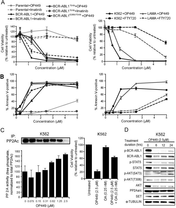
Purpose: The SET oncoprotein, a potent inhibitor of the protein phosphatase 2A (PP2A), is overexpressed in leukemia. We evaluated the efficacy of SET antagonism in chronic myeloid leukemia (CML) and acute myeloid leukemia (AML) cell lines, a murine leukemia model, and primary patient samples using OP449, a specific, cell-penetrating peptide that antagonizes SET's inhibition of PP2A.

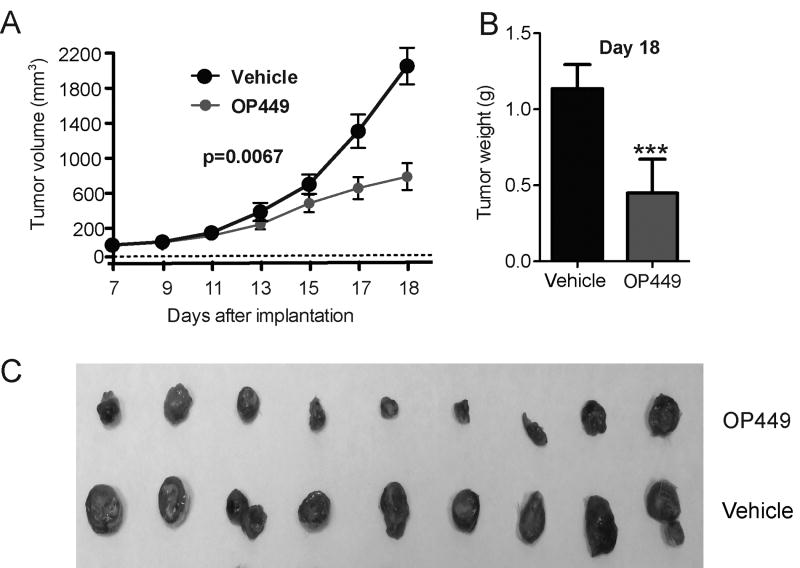
Experimental design: In vitro cytotoxicity and specificity of OP449 in CML and AML cell lines and primary samples were measured using proliferation, apoptosis, and clonogenic assays. Efficacy of target inhibition by OP449 was evaluated by immunoblotting and PP2A assay. In vivo antitumor efficacy of OP449 was measured in human HL-60 xenografted murine model.

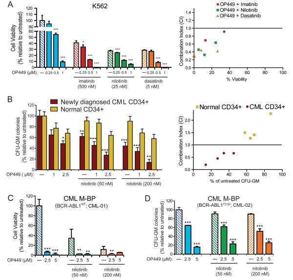
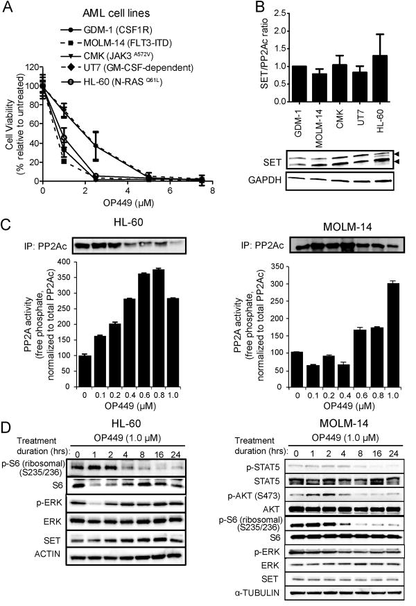
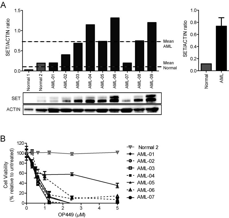
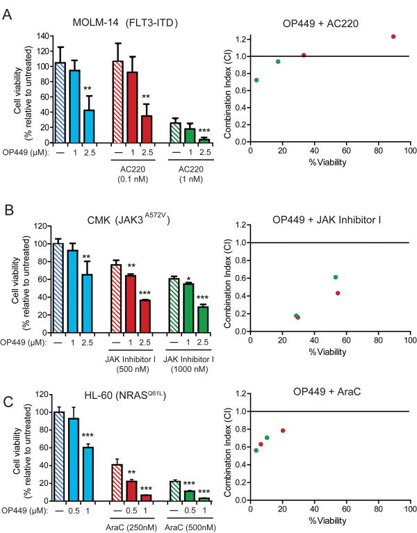
Results: We observed that OP449 inhibited growth of CML cells including those from patients with blastic phase disease and patients harboring highly drug-resistant BCR-ABL1 mutations. Combined treatment with OP449 and ABL1 tyrosine kinase inhibitors was significantly more cytotoxic to K562 cells and primary CD34(+) CML cells. SET protein levels remained unchanged with OP449 treatment, but BCR-ABL1-mediated downstream signaling was significantly inhibited with the degradation of key signaling molecules such as BCR-ABL1, STAT5, and AKT. Similarly, AML cell lines and primary patient samples with various genetic lesions showed inhibition of cell growth after treatment with OP449 alone or in combination with respective kinase inhibitors. Finally, OP449 reduced the tumor burden of mice xenografted with human leukemia cells.

Conclusions: We demonstrate a novel therapeutic paradigm of SET antagonism using OP449 in combination with tyrosine kinase inhibitors for the treatment of CML and AML.


Conflict of interest statement

Disclosure of potential conflicts of interest: B.J.D serves as a consultant to MolecularMD, Blueprint Medicines, Gilead Sciences, Cell Therapeutics, Inc., AstraZeneca, Cylene Pharmaceuticals and Lorus Therapeutics. B.J.D. has a subcontract from Oncotide's NIH STTR 1R41CA165570-01. OHSU has clinical trial contracts with Novartis, Bristol-Myers Squibb and ARIAD to pay for patient costs, nurse and data manager salaries, and institutional overhead. B.J.D. does not derive salary, nor does his laboratory receive funds, from these contracts. OHSU and B.J.D. have a financial interest in Molecular MD. Technology used in some of these studies has been licensed to MolecularMD by OHSU. This potential individual and institutional conflict of interest has been reviewed and managed by OHSU. M.V is an Associate Professor at Duke and his potential individual and institutional conflict of interest has been reviewed and is managed by the DUMC Conflict of Interest Committee. R.P. and M.D.O. declare no competing financial interests. D.J.C. and M.P.V. are shareholders (>5%) and employees of Oncotide Pharmaceuticals.

Figures

Figure 1
Figure 1. OP449 inhibits growth, increases apoptosis, and decreases BCR-ABL1 signaling activity in CML cells by specifically activating PP2A
(A) Effect of OP449 on growth of BCR-ABL1-positive cells. Murine Ba/F3 cells expressing BCR-ABL1, BCR-ABL1T315I or BCR-ABL1E255V/T315I and human CML cells (K562 and LAMA) were cultured in graded concentrations of OP449, imatinib or FTY720 and cell viability was measured at 72 hrs by standard MTS assay. Parental Ba/F3 cells were included as a control. Results are graphed as the mean percent viability relative to untreated cells ± standard deviation. (B) Induction of apoptosis by OP449 in BCR-ABL1-positive cells. Murine Ba/F3, Ba/F3-BCR-ABL1, Ba/F3-BCR-ABL1T315I and Ba/F3-BCR-ABL1E255V/T315I cells and human CML cells (K562 and LAMA) were cultured in graded concentrations of OP449, imatinib or FTY720 and apoptosis was measured at 72 hrs. Results are graphed as the mean percent annexin-positive cells ± standard deviation. (C) Left Panel: Reactivation of PP2A in K562 cells following OP449 treatment. K562 cells were exposed to graded concentrations of OP449 for 24 h and free phosphate levels were measured. Results are presented as free phosphate levels normalized to the total amount of immunoprecipitated PP2Ac protein as detected by western blot analysis± standard deviation. Right Panel: Rescue of OP449-mediated inhibition by okadaic acid. K562 cells were treated with OP449 and okadaic acid (OA) either alone or in combination for 72 hrs and cell viability was measured by standard MTS assay. Bars represent the mean percent viability relative to untreated cells ± standard deviation. (D) Inhibition of BCR-ABL1 signaling by OP449. K562 cells were cultured for the indicated times in the presence of OP449, and whole cell lysates were subjected to SDS-PAGE and immunoblotted using the indicated antibodies. All experiments shown are representative of three independent experiments.
Figure 2
Figure 2. The combination of OP449 with ABL1 tyrosine kinase inhibitors is more effective than single treatments and selective for CML cells over normal cells
(A) Combined effects of OP449 and ABL1 kinase inhibitors on proliferation of CML cells. K562 cells were cultured in the presence of OP449 alone or in combination with imatinib, nilotinib, or dasatinib, and viability was measured at 72 hrs by standard MTS assay. Bars represent the mean percent viability relative to untreated cells ± standard deviation. A combination index (CI) value less than 1.0 indicates drug combinations which have a synergistic effect on inhibition of cell growth. (B) Effect of OP449 in combination with nilotinib on primary CML cells. CD34+ cells isolated from newly diagnosed CML patients (N=4) or healthy donors (N=4) were plated in methocult containing cytokines in the presence of OP449 and nilotinib alone or in combination, and colonies were counted after 2 weeks. Bars represent the mean percent of untreated ± standard deviation. * denotes p<0.05, ** denotes p<0.01 and *** denotes p<0.001, where the effect of treatments on CML CD34+ cells are compared with the respective treatments in normal CD34+ cells. A combination index (CI) value less than 1.0 indicates drug combinations which have a synergistic effect on inhibition of cell growth. (C) Efficacy of OP449 in primary myeloid blastic phase (M-BP) CML samples harboring wild-type BCR-ABL1. Samples were cultured in the presence of OP449 alone or in combination with nilotinib and proliferation was measured at 72 hrs by standard MTS assay. Bars represent the mean percent of untreated ± standard deviation. (D) Efficacy of OP449 in primary ABL1 kinase inhibitor-resistant CML cells. Similar CFU-GM colony assays were performed using CD34+ cells isolated from a CML M-BP patient harboring a BCR-ABL1T315I mutation and treated with OP449 and nilotinib alone and in combination. Bars represent the mean percent of untreated ± standard deviation. For panels A, C, and D, * denotes p<0.05, ** denotes p<0.01 and *** denotes p<0.001 when treatment with OP449 alone was compared to untreated or combination treatment was compared to the respective tyrosine kinase inhibitors alone.
Figure 3
Figure 3. OP449 inhibits growth and downstream kinase signaling in AML cell lines
(A) Inhibition of growth of AML cell lines by OP449. Cells were cultured in the presence of graded concentrations of OP449 for 72 hrs and cell viability was analyzed by standard MTS assay. Results are graphed as the mean percent viability relative to untreated controls ± standard deviation. (B) SET expression levels in AML cells lines tested. SET/ACTIN ratios were quantified from densitometric analysis of protein expression in each of the indicated AML cell lines. (C) Reactivation of PP2A in AML cells following OP449 treatment. HL-60 and MOLM-14 cells were exposed to graded concentrations of OP449 for 24 hrs and free phosphate levels were measured. Results are normalized to the total amount of immunoprecipitated PP2Ac protein as detected by western blot analysis ± standard deviation. (D) Inhibition of downstream kinase signaling in AML cells by OP449. HL-60 and MOLM-14 cells were incubated for the indicated durations in the presence of OP449, harvested and lysed, and immunoblotted with the indicated antibodies. All experiments shown are representative of three independent experiments.
Figure 4
Figure 4. SET is overexpressed in primary AML cells and OP449 inhibits growth of primary AML patient cell
(A) SET expression levels in primary AML samples and normal CD34+ cells tested. SET/ACTIN ratios were quantified from densitometric analysis of protein expression in each of the indicated samples. Dashed lines represent mean SET/ACTIN ratios for normal CD34+ and AML cells, respectively. Bar graph shows mean SET/ACTIN ratios for normal CD34+ and AML cells ± standard error. (B) Inhibition of primary AML cell viability by OP449. Primary AML and normal CD34+ cells were cultured in the presence of graded concentrations of OP449 for 72 hrs and cell viability was analyzed by standard MTS assay. Results are graphed as the mean percent viability relative to untreated controls ± standard deviation.
Figure 5
Figure 5. OP449 inhibits AML tumor growth in vivo
Mice were treated every 3 days with either 5mg/kg OP449 or vehicle control beginning at 7 days post-implantation of HL-60 cells. (A) Inhibition of tumor growth in vivo by OP449. Mean tumor volumes ± standard deviation over the course of treatment are shown. (B) Comparisons of tumor burden between treatments. Final tumor mass for OP449- and vehicle-treated HL-60 tumors harvested on day 18 after implantation was compared. *** denotes p<0.001. (C) Visual representation of extracted tumors is shown.
Figure 6
Figure 6. Combination of OP449 with tyrosine kinase inhibitors or chemotherapy enhances inhibition of AML cell growth
(A) Combined treatment of FLT3-ITD positive AML cells with OP449 and a FLT3 inhibitor. MOLM-14 cells (FLT3-ITD) were incubated for 72 hrs with OP449 and AC220 alone or in combination and cell viability was measured by standard MTS assay. (B) Combined treatment of JAK3-mutant AML cells with OP449 and a pan-JAK inhibitor. CMK cells (JAK3A572V) were incubated for 72 hrs with OP449 and JAK Inhibitor I alone or in combination and cell viability was measured by standard MTS assay. (C) Combined treatment of NRAS-mutant AML cells with OP449 and cytarabine (AraC). HL-60 cells (NRASQ61L) were incubated for 72 hrs with OP449 and AraC alone or in combination and cell viability was measured by standard MTS assay. Results are graphed as the mean percent proliferation relative to untreated controls ± standard deviation. * denotes p<0.05, **denotes p<0.001 and *** denotes p<0.001, where treatment with OP449 alone was compared to untreated or combination treatment was compared to the respective tyrosine kinase inhibitors or chemotherapy alone. Right panels show combination indices (CI) for all drug combinations, where a CI value less than 1.0 is considered synergistic.

Comment in

References

    1. Agarwal A, Byrd J, Deininger MW. The Molecular Biology of the Chronic Leukemias; DeVita, Hellman, and Rosenberg's Cancer: Principles and Practice of Oncology. 2011
    1. Birkenkamp KU, Geugien M, Lemmink HH, Kruijer W, Vellenga E. Regulation of constitutive STAT5 phosphorylation in acute myeloid leukemia blasts. Leukemia : official journal of the Leukemia Society of America, Leukemia Research Fund, UK. 2001;15:1923–31. - PubMed
    1. Druker BJ, Guilhot F, O'Brien SG, Gathmann I, Kantarjian H, Gattermann N, et al. Five-year follow-up of patients receiving imatinib for chronic myeloid leukemia. The New England journal of medicine. 2006;355:2408–17. - PubMed
    1. Chu S, Xu H, Shah NP, Snyder DS, Forman SJ, Sawyers CL, et al. Detection of BCR-ABL kinase mutations in CD34+ cells from chronic myelogenous leukemia patients in complete cytogenetic remission on imatinib mesylate treatment. Blood. 2005;105:2093–8. - PubMed
    1. Traer E, MacKenzie R, Snead J, Agarwal A, Eiring AM, O'Hare T, et al. Blockade of JAK2-mediated extrinsic survival signals restores sensitivity of CML cells to ABL inhibitors. Leukemia : official journal of the Leukemia Society of America, Leukemia Research Fund, UK. 2012;26:1140–3. - PMC - PubMed

Publication types

MeSH terms