

Review
doi: 10.1055/s-0033-1333658.
Management of transcecal renal transplant nephrostomy
Affiliations
- PMID: 24436522
- PMCID: PMC3700794
- DOI: 10.1055/s-0033-1333658
Item in Clipboard
Review
Management of transcecal renal transplant nephrostomy
Semin Intervent Radiol.
2013 Mar.
No abstract available
Figures

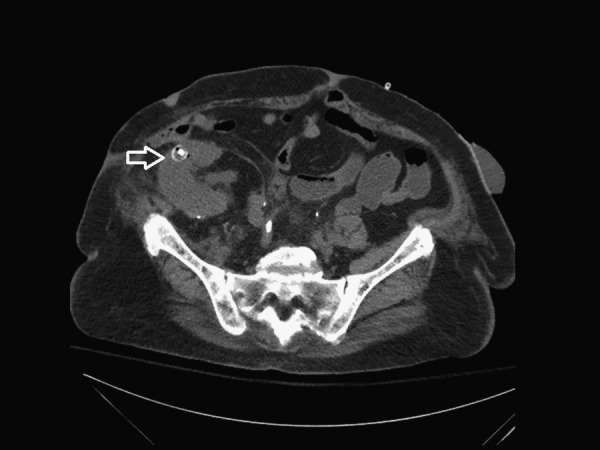
Unenhanced computed tomography demonstrates transcecal placement of indwelling nephrostomy tube with nephroenteric fistula. Solid arrow shows transcecal placement. Open arrow shows pigtail in transplant kidney.
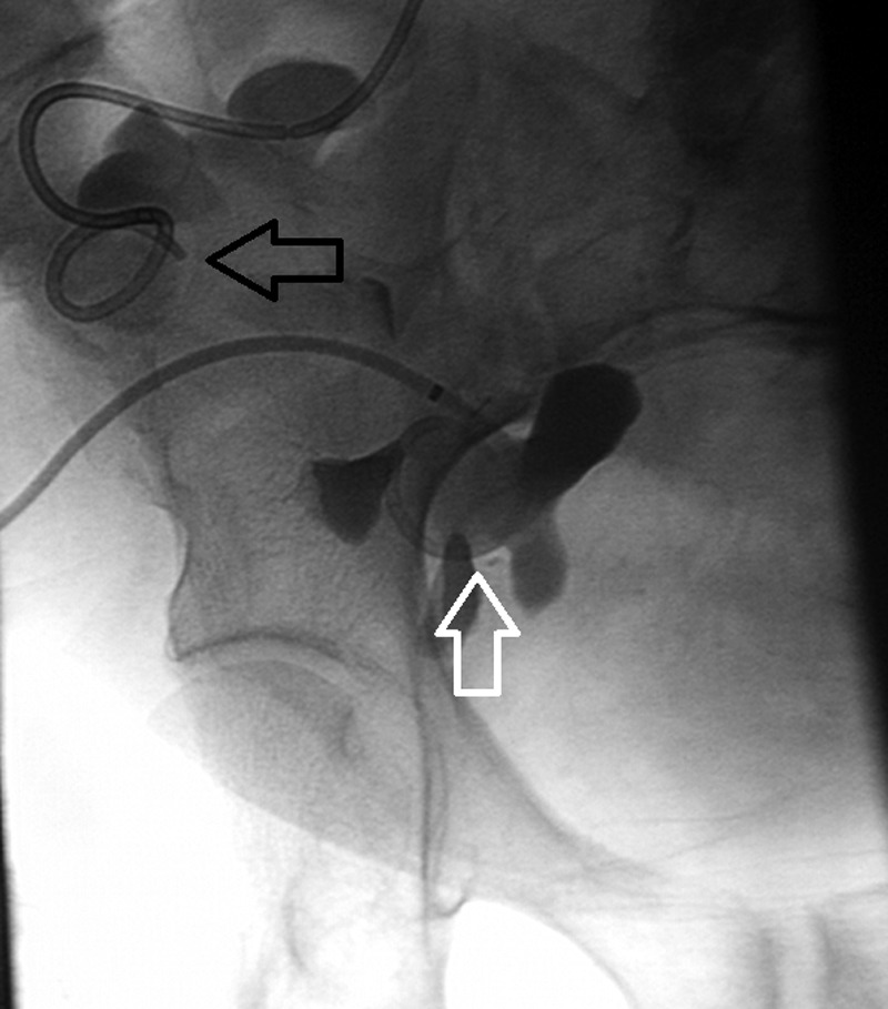
Hydronephrosis with new nephrostomy tube (white arrow) in appropriate position. Black arrow indicates prior transcecal nephrostomy tube.

Tandem wire technique. Tandem guidewires were then placed through the sheath into both the cecum and transplant kidney.

Cecal catheterization while maintaining access to the transplant kidney. Black arrow shows wire in cecum. White arrow shows wire in transplant kidney.

Amplatzer plug deployment across fistula. Arrow shows Amplatzer plug and deployment system.

Final image of cecostomy and nephrostomy tube.

Schematic representation of Amplatzer plug, cecostomy tube, and transplant nephrostomy tubes.

Two-month follow-up examination, following contrast injection via the nephrostomy tube, reveals no communication between the cecostomy (black arrow) and nephrostomy (white arrow) tubes.
Four-month follow-up computed tomography confirms the positioning of the Amplatzer plug (arrow) within the renal parenchyma.
References
-
- Miller G L, Summa J. Transcolonic placement of a percutaneous nephrostomy tube: recognition and treatment. J Vasc Interv Radiol. 1997;8(3):401–403. - PubMed
-
- Saad W EA, Moorthy M, Ginat D. Percutaneous nephrostomy: native and transplanted kidneys. Tech Vasc Interv Radiol. 2009;12(3):172–192. - PubMed
-
- Gerspach J M, Bellman G C, Stoller M L, Fugelso P. Conservative management of colon injury following percutaneous renal surgery. Urology. 1997;49(6):831–836. - PubMed
-
- Zagoria R J, Dyer R B. Do's and don't's of percutaneous nephrostomy. Acad Radiol. 1999;6(6):370–377. - PubMed
Publication types
LinkOut - more resources
Full Text Sources
Other Literature Sources
