Review
. 2013 Dec;30(4):364-71.
doi: 10.1055/s-0033-1359730.

Pelvic vascular malformations

Affiliations
Review

Pelvic vascular malformations

Brian M Christenson et al. Semin Intervent Radiol. 2013 Dec.

Abstract

Vascular malformations (VMs) comprise a wide spectrum of lesions that are classified by content and flow characteristics. These lesions, occurring in both focal and diffuse forms, can involve any organ and tissue plane and can cause significant morbidity in both children and adults. Since treatment strategy depends on the type of malformation, correct diagnosis and classification of a vascular lesion are crucial. Slow-flow VMs (venous and lymphatic malformations) are often treated by sclerotherapy, whereas fast-flow lesions (arteriovenous malformations) are generally managed with embolization. In addition, some cases of VMs are best treated surgically. This review will present an overview of VMs in the female pelvis as well as a discussion of endovascular therapeutic techniques.

Keywords: arteriovenous malformations; endovascular treatment; interventional radiology; lymphatic malformations; vascular malformations; venous malformations.


Figures

Figure 1
Figure 1
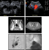
(A) Grayscale ultrasound image demonstrating a sagittal view of the uterus with a hypoechoic cesarian section scar (arrow). (B) Color ultrasound image demonstrating increased vascularity in the region of the cesarian section scar (arrow).
Figure 2
Figure 2
(A) Grayscale ultrasound images demonstrate a hypoechoic lesion in the right adnexa (asterisk). (B) Color ultrasound image demonstrates a vessel (arrow) extending into the lesion with both arterial and venous color Doppler signal characteristics. (C) Coronal CT image demonstrates contrast filling a PSA in the right adnexa (asterisk) with early draining veins (arrow). (D) Axial CT image demonstrates contrast filling a PSA in the right adnexa (asterisk) with early draining veins (arrow). (E) Angiographic image with a base catheter in the proximal uterine artery and a microcatheter advanced into the PSA. Note early venous drainage (arrow). (F) Postembolization angiography via the base catheter in the uterine artery demonstrates multiple coils in the aneurysm sac and feeding uterine artery (arrow). Note no further venous drainage. CT, computed tomography; PSA, pseudoaneurysm.
Figure 3
Figure 3
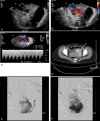
(A) Grayscale ultrasound image demonstrating two large hypoechoic regions (arrows) at the lateral aspect of the uterus. (B) Color ultrasound image demonstrating previous hypoechoic regions have color flow (arrows). (C) Doppler interrogation showing simultaneous arterial and venous waveforms in the uterus. (D) Axial CT image showing contrast in the vascular spaces identified on ultrasound (arrow). A dermoid is incidentally noted (asterisk). (E) Pre-embolization angiogram from the uterine artery (arrow) showing increased vascularity in the left lateral uterus. (F) Delayed phase from previous angiogram demonstrating early venous filling (arrow) and drainage from the uterus. CT, computed tomography.

References

    1. Mulliken J B, Glowacki J. Hemangiomas and vascular malformations in infants and children: a classification based on endothelial characteristics. Plast Reconstr Surg. 1982;69(3):412–422. - PubMed
    1. Jackson I T, Carreño R, Potparic Z, Hussain K. Hemangiomas, vascular malformations, and lymphovenous malformations: classification and methods of treatment. Plast Reconstr Surg. 1993;91(7):1216–1230. - PubMed
    1. Dubois J, Alison M. Vascular anomalies: what a radiologist needs to know. Pediatr Radiol. 2010;40(6):895–905. - PubMed
    1. Moukaddam H, Pollak J, Haims A H. MRI characteristics and classification of peripheral vascular malformations and tumors. Skeletal Radiol. 2009;38(6):535–547. - PubMed
    1. Flors L Leiva-Salinas C Maged I M et al.MR imaging of soft-tissue vascular malformations: diagnosis, classification, and therapy follow-up Radiographics 20113151321–1340., discussion 1340–1341 - PubMed