

Review
doi: 10.1055/s-0032-1332213.
Epub 2013 Jan 16.
Pediatric orbital fractures
Affiliations
- PMID: 24436730
- PMCID: PMC3699236
- DOI: 10.1055/s-0032-1332213
Item in Clipboard
Review
Pediatric orbital fractures
Craniomaxillofac Trauma Reconstr.
2013 Mar.
Abstract
It is wise to recall the dictum "children are not small adults" when managing pediatric orbital fractures. In a child, the craniofacial skeleton undergoes significant changes in size, shape, and proportion as it grows into maturity. Accordingly, the craniomaxillofacial surgeon must select an appropriate treatment strategy that considers both the nature of the injury and the child's stage of growth. The following review will discuss the management of pediatric orbital fractures, with an emphasis on clinically oriented anatomy and development.
Keywords: enophthalmos; entrapment; orbit; pediatric; trauma.
Figures
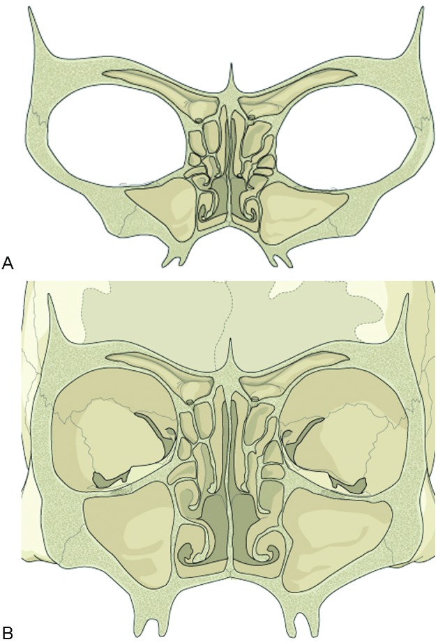
Coronal sections of the pediatric and adult orbit. (A) The thick pediatric orbital floor is contrasted with its diminutive orbital roof. (B) The delicate nature of the adult orbital floor and medial wall is apparent.
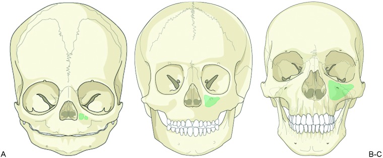
Maxillary sinus development. The progressive increase in size of the maxillary sinus (green) is seen (A) at birth, (B) at 5 years of age, and (C) in the adult.

Cranium-to-face ratio. At birth and in early childhood, the cranium represents a relatively large volume of the craniofacial complex (8:1 and 4:1, respectively). Cranial and orbital fractures are therefore more common during this time. The cranium-to-face ratio in the adult is ∼2:1.
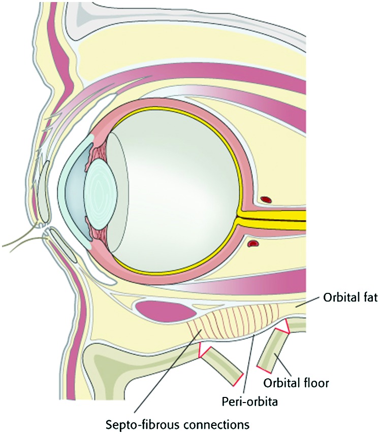
Ligamentous support. The resilient connective tissues of the pediatric orbit allow for expectant management in certain cases of orbital floor fracture. The ligamentous structures prevent expansion of orbital volume.
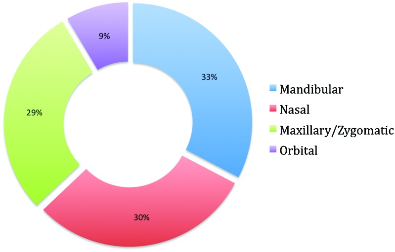
The relative frequency of pediatric facial fractures. In most series of pediatric facial trauma, orbital fractures comprise 5 to 25% of facial fractures. (Adapted from Imahara.)
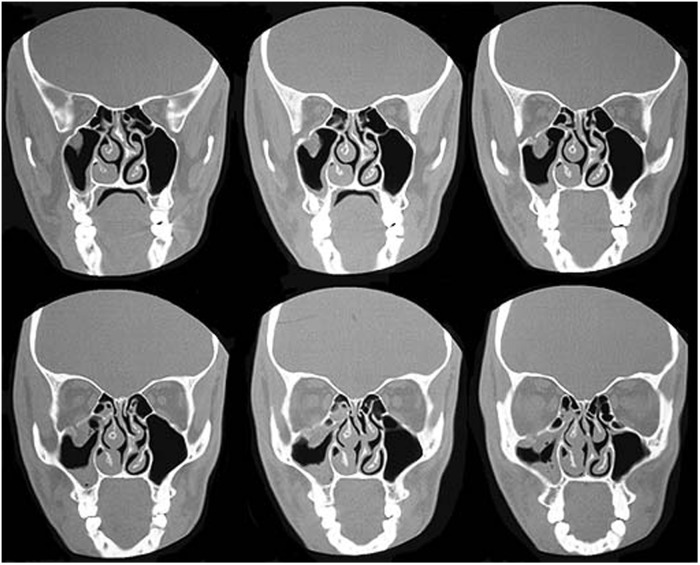
Orbital floor fracture. On this coronal section of a maxillofacial computed tomography scan, orbital contents can be visualized within the maxillary sinus.
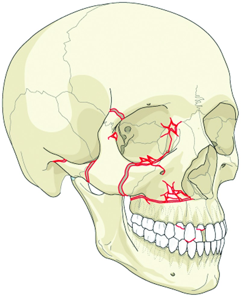
Midface and maxillary fracture patterns.
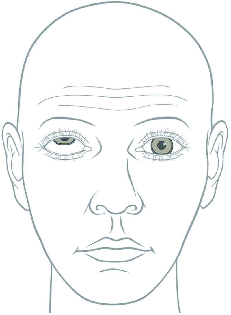
Entrapment. The inferior oblique and/or inferior rectus muscles can become entrapped within an orbital floor fracture. The limitation of extraocular movements can be seen on vertical gaze during physical examination. The patient will experience diplopia during this maneuver. Operative intervention is mandated.
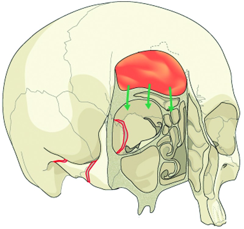
The growing skull fracture. When a dural tear occurs beneath a displaced orbital roof fracture, a leptomeningeal cyst may form during the regenerative process. The cyst interferes with osseous healing, and frontal bone nonunion results. Pulsatile exophthalmos ensues, due to compression (arrows) of the orbital cavity.
References
-
- Enlow D. Philadelphia, PA: WB Saunders; 1982. Handbook of Facial Growth.
-
- Dixon A, Hoyte D A, Rönning O. Salem, MA: CRC Press; 1997. Fundamentals of Craniofacial Growth.
-
- Messinger A Radkowski M A Greenwald M J Pensler J M Orbital roof fractures in the pediatric population Plast Reconstr Surg 198984213–216.; discussion 217–218 - PubMed
-
- Koltai P J, Amjad I, Meyer D, Feustel P J. Orbital fractures in children. Arch Otolaryngol Head Neck Surg. 1995;121:1375–1379. - PubMed
-
- Fortunato M, Manstein G. Facial bone fractures in children. Plast Reconstr Surg. 1982;70:650.
Publication types
LinkOut - more resources
Full Text Sources
Other Literature Sources