Review
. 2013 Jun;3(3):201-18.
doi: 10.1055/s-0033-1350053. Epub 2013 Jul 12.

Notochordal cell-derived therapeutic strategies for discogenic back pain

Affiliations
Review

Notochordal cell-derived therapeutic strategies for discogenic back pain

D Purmessur et al. Global Spine J. 2013 Jun.

Abstract

An understanding of the processes that occur during development of the intervertebral disk can help inform therapeutic strategies for discogenic pain. This article reviews the literature to identify candidates that are found in or derived from the notochord or notochordal cells and evaluates the theory that such factors could be isolated and used as biologics to target the structural disruption, inflammation, and neurovascular ingrowth often associated with discogenic back pain. A systematic review using PubMed was performed with a primary search using keywords "(notochordal OR notochord) And (nerves OR blood vessels OR SHH OR chondroitin sulfate OR notch OR CTGF) NOT chordoma." Secondary searches involved keywords associated with the intervertebral disk and pain. Several potential therapeutic candidates from the notochord and their possible targets were identified. Studies are needed to further identify candidates, explore mechanisms for effect, and to validate the theory that these candidates can promote structural restoration and limit or inhibit neurovascular ingrowth using in vivo studies.

Keywords: CTGF; Notch; Semaphorin 3A; chondroitin sulfate; intervertebral; notochord; notochordal cells; sonic hedgehog.


Conflict of interest statement

Disclosures D. Purmessur, None M. C. Cornejo, None S. K. Cho, None A. C. Hecht, None J. C. Iatridis, None

Figures

Fig. 1
Fig. 1
Schematic of primary and secondary systematic searches used to identify key publications with PubMed as the principal search engine. A primary search of “(notochordal OR notochord) And (nerves OR blood vessels OR SHH OR chondroitin sulfate OR notch OR CTGF) NOT chordoma” identified 390 articles. Of these publications, 37 were considered relevant to the review and were written in the English language. Secondary searches were performed to ensure key areas were not excluded, yielding 49 addition publications.
Fig. 2
Fig. 2
Stages of spine development through maturation and disease. (1) Assembly with the somites alongside the notochord with the sclerotome (orange/pink) situated toward the notochord. (2) Condensation of the sclerotome giving rise to regions of the future intervertebral disk (IVD; orange) and vertebral bodies (VB; pink). (3) Involution of the notochord that expands in the regions of the future IVDs to produce the notochordal cell (NC)-rich nucleus pulposus (NP) and the organized annulus fibrosus (AF) (4). The young NP is highly gelatinous and cellular with large vacuolated NCs. There is distinct demarcation between the NP and AF. (5) The mature IVD transitions with a more fibrous NP region with increased extracellular matrix and decreased cellularity; small nucleus pulposus cells are the predominant cell type in the NP. (6) The degenerate IVD is characterized by a loss of demarcation between the NP and AF, loss in cellularity, structural fissures (white), which provide a path for neurovascular ingrowth (red and brown lines). This figure and concepts are modified from Smith et al, Stemple, and Fleming et al.
Fig. 3
Fig. 3
(A) Stitched image of the sagittal section of degenerated human intervertebral disk (IVD) tissue (Thompson grade IV) stained with FAST demonstrating the regions from which annulus fibrosus (AF), nucleus pulposus (NP), end plate (EP), and Schmorl node (SN) were sampled (circles). (B) Histology of human IVD tissue showing pathologic targets of nerves (top panel), blood vessels (middle panel), and decreased proteoglycan content (bottom panel) colocalized in similar areas of AF, NP, EP, and SN stained with PGP9.5 (Abcam no. ab109261, Cambridge, MA, USA), von Willebrand (Abcam no. ab6994), and FAST (red = collagen, blue = proteoglycan), respectively. Arrows indicate nerves and blood vessels and arrowhead indicates a large capillary network.
Fig. 4
Fig. 4
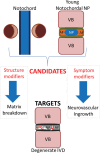
Hypothetical model describing therapeutic candidates and targets for painful disk degeneration. Candidates are identified from the notochord due to their importance in patterning the developing intervertebral disk (IVD) and can be isolated from either the notochord or young notochordal nucleus pulposus (NP). Candidates have structure and symptom-modifying potential to treat targets to limit matrix breakdown and neurovascular ingrowth present in the degenerate IVD. Abbreviations: AF, annulus fibrosus; IVD, intervertebral disk; NP, nucleus pulposus; VB, vertebral bodies.

References

    1. Stemple D L. Structure and function of the notochord: an essential organ for chordate development. Development. 2005;132:2503–2512. - PubMed
    1. Anderson C N, Ohta K, Quick M M, Fleming A, Keynes R, Tannahill D. Molecular analysis of axon repulsion by the notochord. Development. 2003;130:1123–1133. - PubMed
    1. Choi K S, Cohn M J, Harfe B D. Identification of nucleus pulposus precursor cells and notochordal remnants in the mouse: implications for disk degeneration and chordoma formation. Dev Dyn. 2008;237:3953–3958. - PMC - PubMed
    1. Reese D E, Hall C E, Mikawa T. Negative regulation of midline vascular development by the notochord. Dev Cell. 2004;6:699–708. - PubMed
    1. Hunter C J, Matyas J R, Duncan N A. Cytomorphology of notochordal and chondrocytic cells from the nucleus pulposus: a species comparison. J Anat. 2004;205:357–362. - PMC - PubMed

LinkOut - more resources