

Nasopharyngeal branchial cysts-diagnosis and management: a case series
- PMID: 24436888
- PMCID: PMC3699168
- DOI: 10.1055/s-0032-1331020
Nasopharyngeal branchial cysts-diagnosis and management: a case series
Abstract
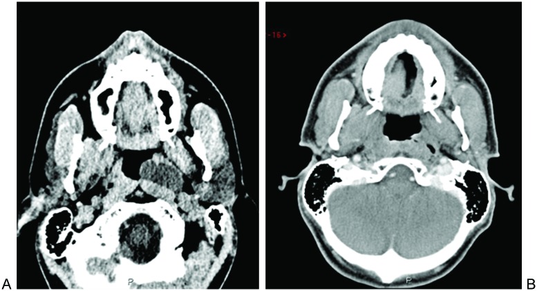
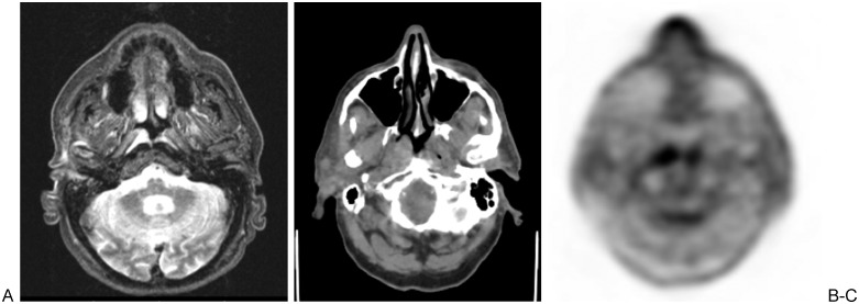
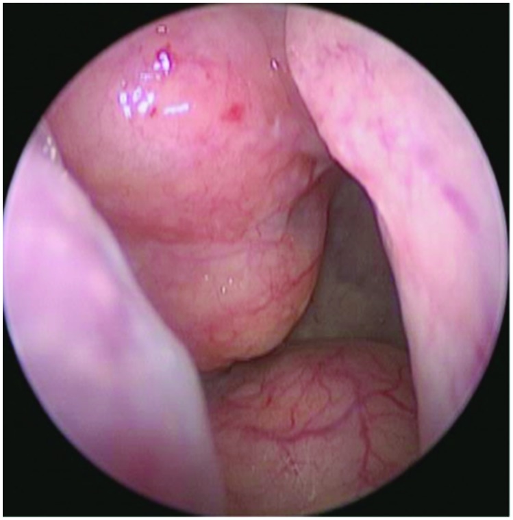
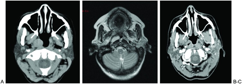
Nasopharyngeal branchial cysts (NBCs) have been discussed in the literature in only a limited number of publications. Differing from Tornwaldt cysts, NBCs present laterally and arise from the fossa of Rosenmuller and may track superiorly within the bony confines of the Eustachian tube. Initially patients are asymptomatic but may present with aural fullness, unilateral conductive hearing loss, and serous otitis media as the cyst mass grows. Two of our three patients had the lesion incidentally identified at the time of assessment for another diagnosis. In this case series, imaging characteristics and response to treatment are reviewed. A literature search was performed to summarize the management options for this entity.
Keywords: Tornwaldt cysts; marsupialization; mucus retention cysts; nasopharyngeal branchial cysts; nasopharyngeal cysts; skull base.
Figures
References
-
- Ali M Y. Pathogenesis of cysts and crypts in the nasopharynx. J Laryngol Otol. 1965;79:391–402. - PubMed
-
- Nicolai P, Luzzago F, Maroldi R, Falchetti M, Antonelli A R. Nasopharyngeal cysts. Report of seven cases with review of the literature. Arch Otolaryngol Head Neck Surg. 1989;115:860–864. - PubMed
-
- Guggenheim P. Cysts of the nasopharynx. Laryngoscope. 1967;77:2147–2168. - PubMed
-
- Ben Salem D, Duvillard C, Assous D, Ballester M, Krausé D, Ricolfi F. Imaging of nasopharyngeal cysts and bursae. Eur Radiol. 2006;16:2249–2258. - PubMed
LinkOut - more resources
Full Text Sources
