. 2014 Apr;155(4):1327-39.
doi: 10.1210/en.2013-1759. Epub 2014 Jan 17.

Liver mTOR controls IGF-I bioavailability by regulation of protein kinase CK2 and IGFBP-1 phosphorylation in fetal growth restriction

Affiliations

Liver mTOR controls IGF-I bioavailability by regulation of protein kinase CK2 and IGFBP-1 phosphorylation in fetal growth restriction

Majida Abu Shehab et al. Endocrinology. 2014 Apr.

Abstract

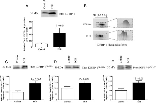
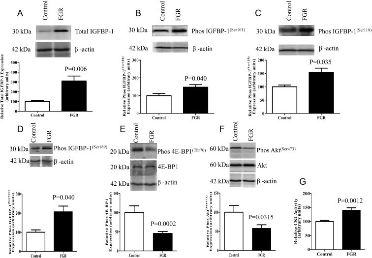
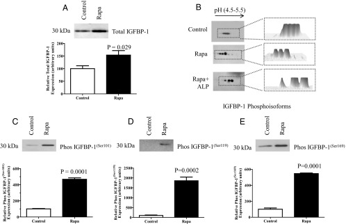
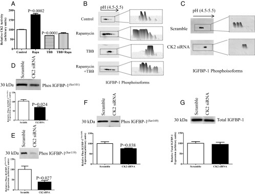
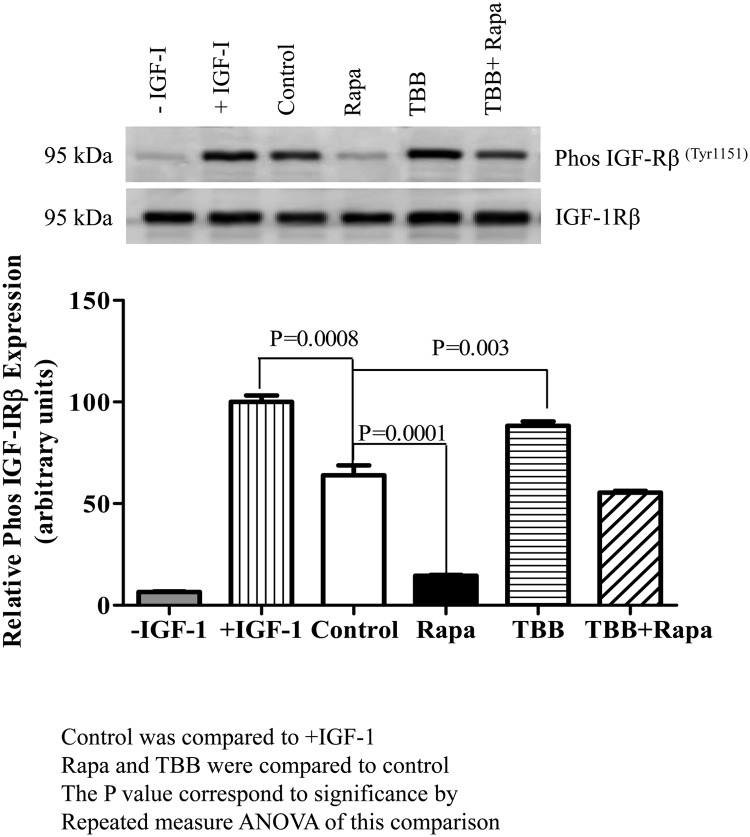
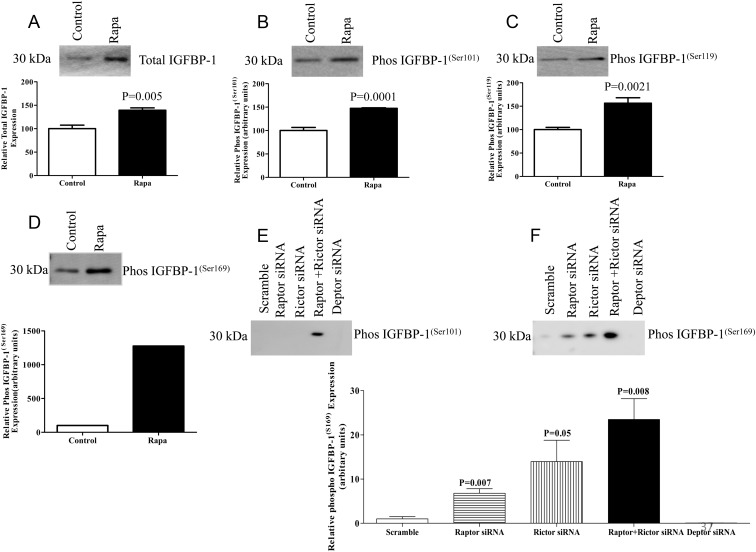
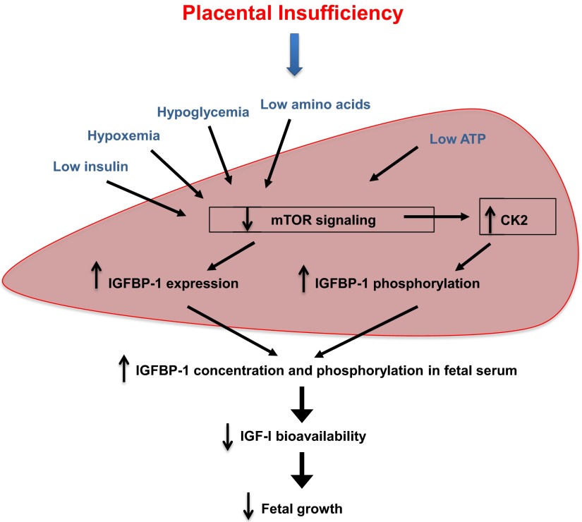
Fetal growth restriction (FGR) increases the risk for perinatal complications and predisposes the infant to diabetes and cardiovascular disease later in life. No treatment for FGR is available, and the underlying pathophysiology remains poorly understood. Increased IGFBP-1 phosphorylation has been implicated as an important mechanism by which fetal growth is reduced. However, to what extent circulating IGFBP-1 is phosphorylated in FGR is unknown, and the molecular mechanisms linking FGR to IGFBP-1 phosphorylation have not been established. We used umbilical cord plasma of appropriate for gestational age (AGA) and growth-restricted human fetuses and determined IGFBP-1 and IGF-I concentrations (ELISA) and site-specific IGFBP-1 phosphorylation (Western blotting using IGFBP-1 phospho-site specific antibodies). In addition, we used a baboon model of FGR produced by 30% maternal nutrient restriction and determined mammalian target of rapamycin (mTOR)C1 activity, CK2 expression/activity, IGFBP-1 expression and phosphorylation, and IGF-I levels in baboon fetal liver by Western blot, enzymatic assay, and ELISA. HepG2 cells and primary fetal baboon hepatocytes were used to explore mechanistic links between mTORC1 signaling and IGFBP-1 phosphorylation. IGFBP-1 was hyperphosphorylated at Ser101, Ser119, and Ser169 in umbilical plasma of human FGR fetuses. IGFBP-1 was also hyperphosphorylated at Ser101, Ser119, and Ser169 in the liver of growth-restricted baboon fetus. mTOR signaling was markedly inhibited, whereas expression and activity of CK2 was increased in growth-restricted baboon fetal liver in vivo. Using HepG2 cells and primary fetal baboon hepatocytes, we established a mechanistic link between mTOR inhibition, CK2 activation, IGFBP-1 hyperphosphorylation, and decreased IGF-I-induced IGF-I receptor autophosphorylation. We provide clear evidence for IGFBP-1 hyperphosphorylation in FGR and identified an mTOR and CK2-mediated mechanism for regulation of IGF-I bioavailability. Our findings are consistent with the model that inhibition of mTOR in the fetal liver, resulting in increased CK2 activity and IGFBP-1 hyperphosphorylation, constitutes a novel mechanistic link between nutrient deprivation and restricted fetal growth.


Figures

Figure 1.
Figure 1.
Total and phosphorylated IGFBP-1 in umbilical cord blood of fetal growth–restricted pregnancies. A, Representative Western blot of total IGFBP-1 in umbilical cord blood of FGR (n = 12) and gestational age–matched AGA controls (n = 12). Equal volumes of plasma samples were loaded. Fetal circulating IGFBP-1 was elevated in human FGR umbilical cord blood. B, Representative 2-D immunoblot of IGFBP-1 phosphoisoforms in umbilical cord blood from control (top) and growth–restricted fetuses (bottom) with a 3-D image analysis of spot intensity (adjacent), using equal aliquots of plasma samples. A marked increase in the intensity of spots and a shift toward the left (positive) was seen, representing increased IGFBP-1 phosphorylation in FGR. C–E, Representative Western blots of IGFBP-1 phosphorylated at Ser101, Ser119, and Ser169 in umbilical cord blood from FGR (n = 12) and gestational age–matched AGA control (n = 12). An equal volume of plasma was used as a loading control. Phosphorylation of circulating fetal IGFBP-1 was increased at the three serine residues in FGR. Values are given as means ± SEM. P < .05 vs control is considered significant as per unpaired Student t test.
Figure 2.
Figure 2.
FGR in baboon fetal liver causes changes to IGFBP-1, mTOR signaling, and CK2 activity. A, Representative Western blot of IGFBP-1 in control (n = 4) and FGR (n = 9) baboon fetal livers. FGR resulted in increased total IGFBP-1 in fetal liver tissue. B–D, Representative Western blots of phosphorylated IGFBP-1 at Ser101, Ser119, and Ser169 in livers from control (n = 4) and growth–restricted fetuses (n = 9). FGR resulted in increased baboon fetal liver IGFBP-1 phosphorylation at the three serine sites. E and F, Representative Western blots of phos 4E-BP1(Thr70) and phos Akt(Ser473) in livers of control (n = 4) and growth–restricted fetuses (n = 9). mTORC1 and mTORC2 signaling was inhibited in FGR fetal liver tissue. G, Liver CK2 activity in control (n = 4) and growth–restricted baboon fetuses (n = 9). The kinase activity assays were performed in triplicate using equal amounts of protein from whole tissue extracts. CK2 activity was enhanced in growth–restricted baboon liver. Values are given as means ± SEM. P ≤ .05 vs control significant as per unpaired Student t test.
Figure 3.
Figure 3.
Effect of rapamycin (Rapa) on IGFBP-1 secretion and phosphorylation in HepG2 cells. A, Representative Western blot of IGFBP-1 secreted by HepG2 cells from control (n = 6) and rapamycin-treated cells (24 hours) (n = 6) using an equal volume of cell medium as a loading control. Rapamycin treatment of HepG2 cells significantly increased IGFBP-1 secretion. B, Representative 2-D Western blot of IGFBP-1 phosphoisoforms in cell medium from control cells (n = 3) and cells treated with rapamycin (n = 3) or treated with rapamycin and alkaline phosphatase (ALP) (n = 3) before 2-D immunoblot analysis as indicated. Equal loading (cell media) was performed. Phosphoisoform spot intensity for each blot is represented as a 3-D image in the adjacent panel. Rapamycin caused a marked increase in the number of spots and a shift toward the left (positive), indicating increased phosphorylation. C–E, Representative Western blots of phosphorylated IGFBP-1 at Ser101, Ser119, and Ser169 in cell media of control (n = 4) and rapamycin-treated cells (n = 4) using equal aliquots of cell media. Rapamycin treatment of HepG2 cells significantly increased IGFBP-1 phosphorylation at 3 serine sites. Values are given as means ± SEM. P < .05 vs control is significant as per unpaired Student t test.
Figure 4.
Figure 4.
Effect of rapamycin (Rapa) and TBB on CK2 activity and IGFBP-1 phosphorylation, and the effect of CK2 siRNA silencing on IGFBP-1 phosphorylation. A, Summary of CK2 activity in HepG2 cell lysate after 24 hours of rapamycin and TBB treatments. The kinase activity assays were performed using equal amounts of protein from whole-cell lysates (n = 3). Rapamycin treatment of HepG2 cells resulted in mTOR inhibition and stimulation of CK2 activity that was prevented by treatment of cells with the CK2 inhibitor TBB. Values are means ± SEM. P < .05 vs control (n = 3) is significant as per Student t test. B, Representative 2-D Western blots (n = 3) provided qualitative assessments of IGFBP-1 phosphoisoforms in HepG2 cell media from control and rapamycin- and TBB-treated cells and from cells treated with both rapamycin and TBB together. An equal volume of cell medium was used as a loading control. Rapamycin treatment increased IGFBP-1 phosphorylation as is indicated by a marked increase in the number of spots and a shift toward the left (positive), representing increased phosphorylation. On the other hand, inhibition of CK2 by TBB attenuated IGFBP-1 phosphorylation induced by rapamycin (n = 3). C, Representative 2-D Western blots of IGFBP-1 phosphoisoforms in the media of HepG2 cells transfected with scramble or CK2 siRNA. The adjacent panels represent the 3-D isoform intensity profile in each sample. IGFBP-1 phosphorylation was markedly decreased in CK2 silenced (α + α′ + β combined) HepG2 cells. D, Representative Western blot of IGFBP-1 (n = 3) secretion in the media of HepG2 cells transfected with scramble or CK2 (α + α′ + β combined) siRNA using equal aliquots of cell media. E–G, Representative Western blots of IGFBP-1 phosphorylation at Ser101, Ser119, and Ser169 in the cell media of HepG2 cells transfected with scramble or CK2 (α + α′ + β combined) siRNA-silenced HepG2 cells showed a decrease in IGFBP-1 phosphorylation at all three phosphorylation sites. Values are given as means ± SEM. P < .05 vs control is significant as per unpaired Student t test.
Figure 5.
Figure 5.
Effect of IGFBP-1 phosphorylation on IGF-I–induced IGF-IR autophosphorylation. A representative Western blot of phospho IGF-IRβ in P6 cells after incubation with HepG2 cell media consisting of an equal concentration (100 ng) of total IGFBP-1 is shown. An immunoenzymometric assay (Medix Biochemica) was performed for the quantitative determination of IGFBP-1 as per the manufacturer's protocol. Samples from control (no treatment) and from HepG2 cells treated with rapamycin (Rapa), TBB, or TBB + rapamycin combined were preincubated with IGF-I. Rapamycin caused a pronounced inhibition of IGF-I–induced IGF-IR autophosphorylation in P6 cells, whereas TBB stimulated IGF-IRβ autophosphorylation, and TBB + rapamycin had no significant effect on IGF-IR activity compared with that in control cells (no treatment) as indicated. The comparison between treatments was done by Repeated Measures ANOVA and the Bonferroni multiple comparison test.
Figure 6.
Figure 6.
Effect of rapamycin (Rapa) on IGFBP-1 expression and phosphorylation, and mTORC1 and mTORC2 siRNA silencing on IGFBP-1 phosphorylation in fetal baboon hepatocytes. A, Representative Western blot of IGFBP-1 expression in baboon fetal hepatocytes treated with rapamycin (25 nM as indicated) (n = 3). Equal loading was performed. B–D, Representative Western blots of phosphorylated IGFBP-1 at Ser101, Ser119, and Ser169 in baboon fetal hepatocytes treated with rapamycin (n = 3). Equal loading was performed. E and F, Representative Western blots of IGFBP-1 phosphorylation at Ser101 and Ser169 in cell media of baboon hepatocytes transfected with scramble, rictor, raptor, rictor + raptor, and DEPTOR siRNA (n = 3 each). Values are given as means ± SEM. P < .05 vs control is significant as per unpaired Student t test. Rapamycin treatment (25 nm) of primary fetal baboon hepatocytes in 24 hours significantly enhanced IGFBP-1 secretion and phosphorylation.
Figure 7.
Figure 7.
The proposed model. Inhibition of mTOR signaling and activation of protein kinase CK2 in the fetal liver constitutes a key molecular link between nutrient deprivation, increased IGFBP-1 secretion and phosphorylation, and decreased IGF-I bioavailability in FGR.

References

    1. Pallotto EK, Kilbride HW. Perinatal outcome and later implications of intrauterine growth restriction. Clin Obstet Gynecol. 2006;49:257–269 - PubMed
    1. Gluckman PD, Hanson MA, Cooper C, Thornburg KL. Effect of in utero and early-life conditions on adult health and disease. N Engl J Med. 2008;359:61–73 - PMC - PubMed
    1. Liu JP, Baker J, Perkins AS, Robertson EJ, Efstratiadis A. Mice carrying null mutations of the genes encoding insulin-like growth factor I (Igf-1) and type 1 IGF receptor (Igf1r). Cell. 1993;75:59–72 - PubMed
    1. Lassarre C, Hardouin S, Daffos F, Forestier F, Frankenne F, Binoux M. Serum insulin-like growth factors and insulin-like growth factor binding proteins in the human fetus. Relationships with growth in normal subjects and in subjects with intrauterine growth retardation. Pediatr Res. 1991;29:219–225 - PubMed
    1. Gibson JM, Aplin JD, White A, Westwood M. Regulation of IGF bioavailability in pregnancy. Mol Hum Reprod. 2001;7:79–87 - PubMed

Publication types

MeSH terms

Substances