

Reconstruction of an active SOCS3-based E3 ubiquitin ligase complex in vitro: identification of the active components and JAK2 and gp130 as substrates
- PMID: 24438103
- PMCID: PMC4085236
- DOI: 10.3109/08977194.2013.877005
Reconstruction of an active SOCS3-based E3 ubiquitin ligase complex in vitro: identification of the active components and JAK2 and gp130 as substrates
Abstract
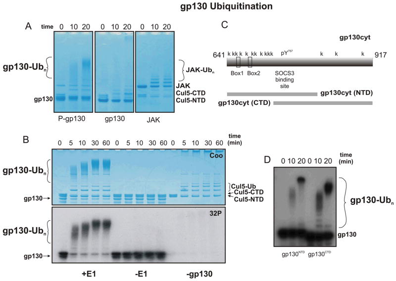
SOCS3 (suppressor of cytokine signaling 3) inhibits the intracellular signaling cascade initiated by exposure of cells to cytokines. SOCS3 regulates signaling via two distinct mechanisms: directly inhibiting the catalytic activity of Janus kinases (JAKs) that initiate the intracellular signaling cascade and catalysing the ubiquitination of signaling components by recruiting components of an E3 ubiquitin ligase complex. Here we investigate the latter mode-of-action biochemically by reconstructing a SOCS3-based E3 ubiquitin ligase complex in vitro using fully purified, recombinant components and examining its ability to promote the ubiquitination of molecules involved in the cytokine signaling cascade. We show that SOCS3 is an active substrate recruitment module for a Cullin5-based E3 ligase and have defined the core protein components required for ubiquitination. SOCS3-induced polyubiquitination was rapid and could proceed through a number of different ubiquitin lysines. SOCS3 catalyzed the ubiquitination of both the IL-6 receptor common chain (gp130) and JAK2.
Conflict of interest statement
NAN is a patent holder on SOCS3 and its activities. This work was made possible through Victorian State Government Operational Infrastructure Support and Australian Government NHMRC IRIISS. This research was supported by an NHMRC Program Grant (461219), NIH Grant (CA022556), ARC Future Fellowship (FT110100169) and NHMRC Project Grant (1011804). The authors alone are responsible for the content and writing of the paper.
Figures
Similar articles
-
Biophysical studies on interactions and assembly of full-size E3 ubiquitin ligase: suppressor of cytokine signaling 2 (SOCS2)-elongin BC-cullin 5-ring box protein 2 (RBX2).J Biol Chem. 2015 Feb 13;290(7):4178-91. doi: 10.1074/jbc.M114.616664. Epub 2014 Dec 11. J Biol Chem. 2015. PMID: 25505247 Free PMC article.
-
The SOCS box domain of SOCS3: structure and interaction with the elonginBC-cullin5 ubiquitin ligase.J Mol Biol. 2008 Sep 12;381(4):928-40. doi: 10.1016/j.jmb.2008.06.038. Epub 2008 Jun 20. J Mol Biol. 2008. PMID: 18590740 Free PMC article.
-
The SOCS box encodes a hierarchy of affinities for Cullin5: implications for ubiquitin ligase formation and cytokine signalling suppression.J Mol Biol. 2009 Mar 20;387(1):162-74. doi: 10.1016/j.jmb.2009.01.024. J Mol Biol. 2009. PMID: 19385048 Free PMC article.
-
Role of SOCS and VHL Proteins in Neuronal Differentiation and Development.Int J Mol Sci. 2023 Feb 15;24(4):3880. doi: 10.3390/ijms24043880. Int J Mol Sci. 2023. PMID: 36835292 Free PMC article. Review.
-
Inhibition of IL-6 family cytokines by SOCS3.Semin Immunol. 2014 Feb;26(1):13-9. doi: 10.1016/j.smim.2013.12.004. Epub 2014 Jan 10. Semin Immunol. 2014. PMID: 24418198 Free PMC article. Review.
Cited by
-
Structural Understanding of Interleukin 6 Family Cytokine Signaling and Targeted Therapies: Focus on Interleukin 11.Front Immunol. 2020 Jul 16;11:1424. doi: 10.3389/fimmu.2020.01424. eCollection 2020. Front Immunol. 2020. PMID: 32765502 Free PMC article. Review.
-
Leukaemia inhibitory factor in gastric cancer: friend or foe?Gastric Cancer. 2022 Mar;25(2):299-305. doi: 10.1007/s10120-022-01278-w. Epub 2022 Feb 2. Gastric Cancer. 2022. PMID: 35106710 Review.
-
Nicotine aggravates vascular adiponectin resistance via ubiquitin-mediated adiponectin receptor degradation in diabetic Apolipoprotein E knockout mouse.Cell Death Dis. 2021 May 18;12(6):508. doi: 10.1038/s41419-021-03772-y. Cell Death Dis. 2021. PMID: 34006831 Free PMC article.
-
Promotion of Erythropoietic Differentiation in Hematopoietic Stem Cells by SOCS3 Knock-Down.PLoS One. 2015 Aug 7;10(8):e0135259. doi: 10.1371/journal.pone.0135259. eCollection 2015. PLoS One. 2015. PMID: 26252772 Free PMC article.
-
Host and Viral Proteins Modulating Ebola and Marburg Virus Egress.Viruses. 2019 Jan 3;11(1):25. doi: 10.3390/v11010025. Viruses. 2019. PMID: 30609802 Free PMC article. Review.
References
-
- Hilton DJ, Richardson RT, Alexander WS, Viney EM, Willson TA, Sprigg NS, Starr R, Nicholson SE, Metcalf D, Nicola NA. Twenty proteins containing a C-terminal SOCS box form five structural classes. Proceedings of the National Academy of Sciences of the United States of America. 1998;95:114–119. - PMC - PubMed
-
- Sasaki A, Yasukawa H, Suzuki A, Kamizono S, Syoda T, Kinjyo I, Sasaki M, Johnston JA, Yoshimura A. Cytokine-inducible SH2 protein-3 (CIS3/SOCS3) inhibits Janus tyrosine kinase by binding through the N-terminal kinase inhibitory region as well as SH2 domain. Genes to Cells. 1999;4:339–351. - PubMed
Publication types
MeSH terms
Substances
Grants and funding
LinkOut - more resources
Full Text Sources
Other Literature Sources
Miscellaneous