Case Reports
. 2014 Jan 20:12:17.
doi: 10.1186/1477-7819-12-17.

Response of an aggressive periosteal aneurysmal bone cyst (ABC) of the radius to denosumab therapy

Affiliations
Case Reports

Response of an aggressive periosteal aneurysmal bone cyst (ABC) of the radius to denosumab therapy

Chantal Pauli et al. World J Surg Oncol. .

Abstract

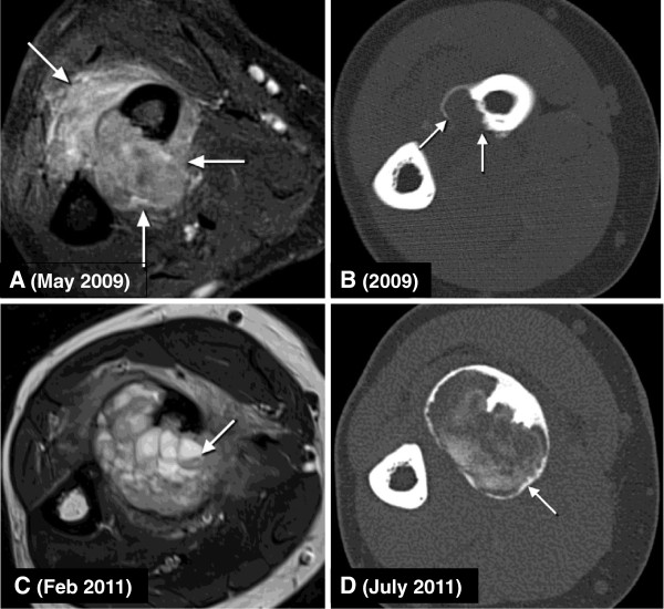
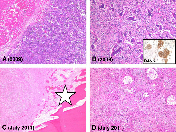
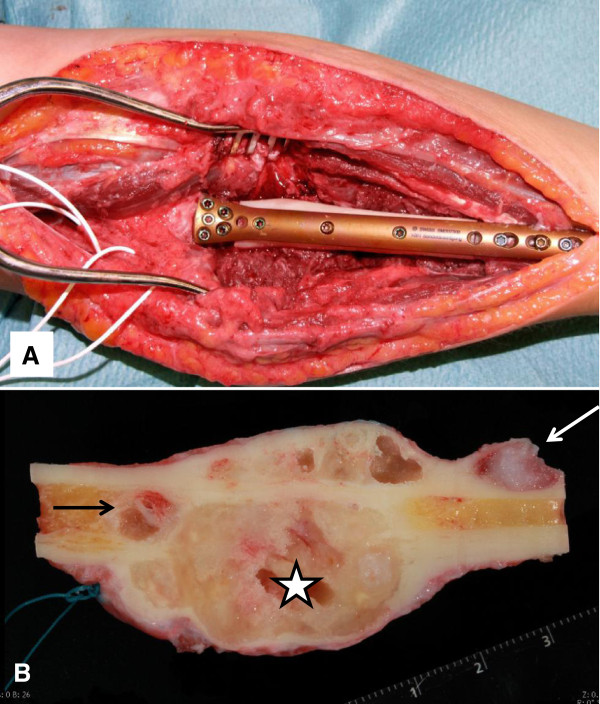
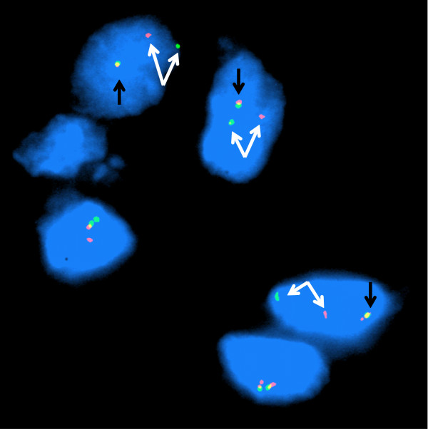
Aneurysmal bone cyst (ABC), once considered a reactive lesion, has been proven to be a neoplasia characterized by rearrangements of the USP6-gene. Aggressive local growth and recurrences are common and therapeutic options may be limited due to the vicinity of crucial structures. We describe a case of a locally aggressive, multinucleated giant cell-containing lesion of the forearm of a 21-year old woman, treated with denosumab for recurrent, surgically uncontrollable disease. Under the influence of this RANKL inhibitor, the tumor showed a marked reduction of the content of the osteoclastic giant cells and an extensive metaplastic osteoid production leading to the bony containment, mostly located intracortically in the proximal radius. The diagnosis of a periosteal ABC was confirmed by FISH demonstrating USP6 gene rearrangement on the initial biopsy. Function conserving surgery could be performed, enabling reconstruction of the affected bone. Inhibition of RANKL with denosumab may offer therapeutic option for patients not only with giant cell tumors but also with ABCs.


Figures

Figure 1
Figure 1
Imaging of the patients right forearm tumor. (A) Initial magnetic resonance imaging (MRI) demonstrating extensive involvement of the soft tissue between the radius and ulna as well as the cortex of the radius by an exclusively solid tumor mass (arrows). (B) Pre-treatment computer tomography (CT) scan with a small area of a split and disrupted cortex of the radius (arrows). (C) MRI directly prior to denosumab therapy with a locally progressive, extensive soft tissue mass following local surgical therapy 18 months previously. Fluid-fluid levels may be seen at this point (arrow). (D) CT scan following five months of denosumab therapy demonstrating almost complete containment of the soft tissue mass by a boney rim (arrow).
Figure 2
Figure 2
Histopathology of the pre- and post-treatment tumor tissue specimen. (A) Pre-treatment biopsy sample showing giant cell containing soft tissue mass with extensive infiltration of the skeletal muscle (H&E stain; original magnification 50×). (B) Abundant lesional giant cells with numerous nuclei and mononuclear cells in the background (H&E stain; original magnification 100×). Inset shows immunohistochemical expression of RANK (dilution 1:400; R&D Systems, Abingdon, United Kingdom) by the osteoclastic giant cells. (C) Denosumab treatment induced boney containment (asterisk) of the tumor (upper left) (H&E stain; original magnification 25×). (D) Post-denosumab-treatment tumor specimen showing pronounced reduction of the number of giant cells (H&E stain; original magnification 100×).
Figure 3
Figure 3
Resection of the shaft of the proximal radius following the denosumab therapy. (A) Reconstruction using an intercalary fibula-allograft and a custom-made plate. (B) Longitudinal section of the resected specimen demonstrating large intracortical, fully contained by a rim of bone tumor mass (asterisk) with a small intramedullar tumor nodule proximally (black arrow) and an intracortical satellite distally (white arrow).
Figure 4
Figure 4
Fluorescence in situ hybridization conducted on the pretreatment specimen with a custom-designed break apart probe set; the probe cocktail proximal to the USP6 locus is labeled in green and distal in red. The mononuclear nuclei exhibit one fused red/green signal corresponding to a normal 17p USP6 locus (black arrow) and a pair of split green and red signals (white arrows) indicating a rearrangement of the USP6 locus consistent with the diagnosis of aneurysmal bone cyst.

References

    1. Campanacci M, Cervellati C, Donati U, Bertoni F. Aneurysmal bone cyst (a study of 127 cases, 72 with long term follow up) Ital J Orthop Traumatol. 1976;2:341–353. - PubMed
    1. Mankin HJ, Hornicek FJ, Ortiz-Cruz E, Villafuerte J, Gebhardt MC. Aneurysmal bone cyst: a review of 150 patients. J Clin Oncol. 2005;23:6756–6762. doi: 10.1200/JCO.2005.15.255. - DOI - PubMed
    1. Cottalorda J, Kohler R, Sales de Gauzy J, Chotel F, Mazda K, Lefort G, Louahem D, Bourelle S, Dimeglio A. Epidemiology of aneurysmal bone cyst in children: a multicenter study and literature review. J Pediatr Orthop B. 2004;13:389–394. doi: 10.1097/01202412-200411000-00008. - DOI - PubMed
    1. Lichtenstein L. Aneurysmal bone cyst; further observations. Cancer. 1953;6:1228–1237. doi: 10.1002/1097-0142(195311)6:6<1228::AID-CNCR2820060617>3.0.CO;2-A. - DOI - PubMed
    1. Oliveira AM, Hsi BL, Weremowicz S, Rosenberg AE, Dal Cin P, Joseph N, Bridge JA, Perez-Atayde AR, Fletcher JA. USP6 (Tre2) fusion oncogenes in aneurysmal bone cyst. Cancer Res. 2004;64:1920–1923. doi: 10.1158/0008-5472.CAN-03-2827. - DOI - PubMed

Publication types

MeSH terms