. 2014 Apr;224(4):432-46.
doi: 10.1111/joa.12159. Epub 2014 Jan 18.

Anatomy of the lamprey ear: morphological evidence for occurrence of horizontal semicircular ducts in the labyrinth of Petromyzon marinus

Affiliations

Anatomy of the lamprey ear: morphological evidence for occurrence of horizontal semicircular ducts in the labyrinth of Petromyzon marinus

Adel Maklad et al. J Anat. 2014 Apr.

Abstract

In jawed (gnathostome) vertebrates, the inner ears have three semicircular canals arranged orthogonally in the three Cartesian planes: one horizontal (lateral) and two vertical canals. They function as detectors for angular acceleration in their respective planes. Living jawless craniates, cyclostomes (hagfish and lamprey) and their fossil records seemingly lack a lateral horizontal canal. The jawless vertebrate hagfish inner ear is described as a torus or doughnut, having one vertical canal, and the jawless vertebrate lamprey having two. These observations on the anatomy of the cyclostome (jawless vertebrate) inner ear have been unchallenged for over a century, and the question of how these jawless vertebrates perceive angular acceleration in the yaw (horizontal) planes has remained open. To provide an answer to this open question we reevaluated the anatomy of the inner ear in the lamprey, using stereoscopic dissection and scanning electron microscopy. The present study reveals a novel observation: the lamprey has two horizontal semicircular ducts in each labyrinth. Furthermore, the horizontal ducts in the lamprey, in contrast to those of jawed vertebrates, are located on the medial surface in the labyrinth rather than on the lateral surface. Our data on the lamprey horizontal duct suggest that the appearance of the horizontal canal characteristic of gnathostomes (lateral) and lampreys (medial) are mutually exclusive and indicate a parallel evolution of both systems, one in cyclostomes and one in gnathostome ancestors.

Keywords: horizontal ducts; jawless vertebrates; lamprey; parallel evolution.


Figures

Figure 1
Figure 1
Location and orientation of the labyrinth in lamprey. (A) Lateral surface of the skull, with otic capsule exposed, showing the oval shape of the otic capsule (OC) with the long, horizontal axis (dotted line a) passing 1 cm below the dorsal midline (dotted line c), which runs through the dorsal opening of nasohypophyseal organ (NHO). The short vertical axis (dotted line b) passes approximately 7 mm behind the center of the eye. (B) Lateral surface of the membranous labyrinth after removing the lateral wall of the otic capsule. Note that the membranous labyrinth fills the entire otic capsule. (C) Medial surface of the otic capsule (OC) viewed from the interior of skull. The horizontal axis passes through the upper margin of the optic canal (thick arrow), which is located at the anterior pole of otic capsule. The opening for the VIII cranial nerve (thin arrow) is located at the intersection of horizontal and vertical axes (dotted lines a and b). The opening for the endolymphatic duct (arrowhead) lies at the upper margin of the otic capsule at the vertical axis. (D) The medial surface of the membranous labyrinth after removal of the medial wall of the otic capsule. On the medial wall, the mean plane of two horizontal ducts is located at the horizontal axis, the equator (dotted line a). The mean planes for the two vertical ducts (dotted lines c and d) are located on the medial surface on diagonal planes midway between the horizontal and vertical axes (line a and b).
Figure 2
Figure 2
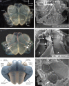
The anatomical features on the lateral wall of the membranous labyrinth in the lamprey. (A, B) Lateral views of the membranous labyrinth of the left ear showing the anterior and posterior ciliated chambers (ACCh and PCCh, respectively, in A). The anterior and the posterior lateral ducts (ALD and PLD, respectively in A; traced in red dotted lines in B) run on the lateral surfaces of the anterior and posterior ciliated chambers, roughly at the equator. At the anterior pole, the anterior lateral duct widens to form the anterior common ampulla (ACA in A) from which two anterior ducts start: the anterior horizontal duct and anterior vertical duct (AHD and AVD, respectively, in A). Their openings are traced in green and blue, respectively, in B. In the ampulla, there is a two-armed crista (traced in black in B); the vertical arm (thin arrow) sits at the opening of the horizontal duct and the horizontal arm (thick arrow) at the opening of the vertical duct. At the divide between the two ducts is the cruciate eminence (arrowhead in B). Similarly, at the posterior pole, the posterior lateral duct widens to form the posterior common ampulla (PCA in A) from which the posterior horizontal and posterior vertical ducts start (PHD and PVD, respectively, in A). (C) Lateral surface of the labyrinth in a 3D reconstruction of the labyrinth. (D) SEM image of the lateral surface of the labyrinth after removing the lateral wall of the ALD and PLD. The upper parts of the lateral ducts are adjacent to each other but are separated by a wall (thin arrows) which is reinforced medially by the spindle organ (thick arrow). The two lateral ducts open superiorly via a single common opening (stars) into the ciliated chambers. Inferiorly, each lateral duct bifurcates at the ampulla into two ducts: the horizontal and vertical ducts. The lateral openings of the ducts into the ampullae are indicated by dotted circles. The interior of the lateral ducts shows a prominent ridge on the medial wall (arrowheads). (E) A higher magnification of the upper area in (D). (F) A higher magnification image of the lower, ampullary region in (D), showing the lateral openings of the horizontal and vertical ducts into the ampullae (dotted circles). The vertical crista (thin arrow) sits at the horizontal duct opening and the horizontal crista (thick arrow) at the opening of the vertical duct. The two cristae are separated by the cruciate eminence (arrowhead), which extends medially to form the partition between the horizontal and vertical ducts proper (stars).
Figure 3
Figure 3
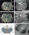
The anatomical features on the medial wall of the membranous labyrinth in the lamprey. (A, B) Stereoscopic dissection images of the medial wall of labyrinth, showing anterior and posterior ciliated chambers (ACCh and PCCh in A). The anterior and posterior horizontal ducts proper (AHD and PHD in A; traced in green dotted lines in B) are shown passing in the plane of the equator (dotted horizontal line in B). The medial wall also has the two vertical duct pairs: the anterior and posterior vertical ducts proper (AVD and PVD in A; traced in blue dotted lines in B). The vertical ducts proper pass in the diagonal planes (dotted diagonal line). A bifid crista (traced in black dotted line in B) sits at the ampullary ends of the ducts. The vertical crista (thin arrow in A) sits at the ampullary end of the horizontal duct, whereas the horizontal crista (thick arrow in A) sits at the ampullary ends of the vertical duct. The cruciate eminence between the two cristae (arrowhead in A) is level with the partition between the two ducts. (C) A 3D reconstruction of the medial wall of the labyrinth. (D) SEM image of a medial view of the medial wall of the labyrinth that shows the horizontal and vertical duct pairs (traced in green and blue dotted lines, respectively) after removing their medial walls to show their luminal surfaces. The horizontal and vertical duct pairs are separated by a thick wall (stars). (E) SEM image of a lateral view of the medial surface as examined from the inside of the ciliated chambers. The openings of the horizontal ducts (green dotted triangles) and the the vertical ducts (blue dotted triangles) are shown close to the midline partition between the ciliated chambers (dotted line). Between the openings of the PHD and PVD is the opening of the saccular pouch (traced in white dotted lines). The wall separating the horizontal and vertical ducts extend beyond their openings to form the anterior and posterior horizontal maculae (arrows and arrowheads, respectively). (F) A higher magnification of the PHD and PVD openings and posterior horizontal macula (arrowheads).
Figure 4
Figure 4
The anatomical features on the dorsal view of the membranous labyrinth in the lamprey. (A, B) Stereoscopic dissection images of the dorsal view of the membranous labyrinth. The dorsal surfaces of the anterior and posterior ciliated chambers (ACCh and PCCh in A) are shown. The anterior and posterior lateral ducts (ALD and PLD in A; traced in red dotted line in B) wrap around the lateral surfaces of ciliated chambers. The anterior and posterior horizontal ducts proper (AHD and PHD in A; traced in green dotted lines in B) wrap around the medial surfaces of their respective ciliated chambers. Each horizontal duct proper and its respective lateral duct form a complete semicircle flanking their respective ciliated chambers. The anterior and posterior common ampullae (ACA and PCA in A) are at the peak of their respective semicircular ducts. (C) A 3D reconstruction of the anatomical features on the dorsal view of the labyrinth. (D) SEM image of the ventromedial quadrant of the membranous labyrinth in which the lateral walls of the ducts proper have been removed to examine their lumens. The lumen of the horizontal duct is traced in green and that of the vertical duct in blue. At the ampullary end of the horizontal duct proper was the vertical crista (thin arrow) and at the ampullary end of the vertical duct proper is the horizontal crista (thick arrow). The cruciate eminence (arrowhead) is continuous with the thick septum separating the horizontal and vertical ducts (stars). (E) A higher magnification of the anterior half of D.
Figure 5
Figure 5
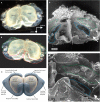
The anatomical features on the ventral view of the labyrinth of the lamprey. (A, B) Stereoscopic dissection images of the ventral view of the membranous labyrinth. This view shows the narrow ventral surfaces of the anterior and posterior ciliated chambers (ACCh and PCCh in A) and the anterior and posterior lateral ducts (ALD and PLD in A; traced in red dotted line in B). In the ventral view, the entire course of the anterior and posterior vertical ducts proper is shown (AVD and PVD in A; traced in blue dotted line in B). Each lateral duct, with its respective vertical duct proper, forms a complete semicircle with an ampulla on its peak (ACA and PCA in A). The ventral surfaces of the horizontal cristae are shown (arrows in A; traced in black dotted lines in B). The vertical orientation of the vertical ducts allow the lateral outer margins of the horizontal ducts to be seen from the ventral view (traced in green dotted line in B). (C) A 3D reconstruction of the anatomical features as viewed from the ventral aspect of the labyrinth.
Figure 6
Figure 6
A model of the role of the horizontal ducts in postural control of movement in the yaw plane. When the animal turns its head to the right, the right posterior and left anterior horizontal ducts are activated (+), whereas the right anterior and left posterior horizontal ducts are deactivated (−). Connectivity patterns of the direct and crossed vestibuloreticular neurons (VRNs) direct the facilitated inputs to reticulospinal neurons (RSNs) projecting to the ventral and dorsal spinal motor neurons on the left side of the body, resulting in a corrective leftward body turn. The minor direct and crossed inhibitory pathways (neurons drawn in gray lines) end as disinhibition on the left side and facilitated inhibition on the right side, further augmenting the effect of the major pathways.
Figure 7
Figure 7
A model of the role of the horizontal ducts in vestibuloocular reflexes to head rotation in the yaw plane. The secondary vestibular neurons in the octavomotorius (OMNs) project equally bilaterally and directly to motor neurons supplying the extraocular muscles. When the animal turns its head to the right, the right posterior and left anterior horizontal ducts are activated (+), whereas the right anterior and left posterior horizontal ducts are deactivated (−). Afferent information from the activated ducts, carried via the left posterior and right anterior divisions of the vestibular nerve (right pVIII and left aVIII), is projected to the right rostral rectus (RR) and left caudal rectus (C.R.) motor neurons via secondary vestibular neurons in the octavomotorius, generating horizontal rostral and caudal movements in the right and left eyes, respectively. There is no evidence that afferent information from the deactivated ducts has any effect on the antagonistic groups of muscles, i.e. right caudal rectus (C.R.) and left rostral rectus (RR), or any evidence of the presence of inhibitory interneurons between the motor neuron nuclei supplying antagonistic groups of muscle.

Similar articles

Cited by

References

    1. Brodin L, Grillner S, Dubuc R. Reticulospinal neurons in lamprey: transmitters, synaptic interactions and their role during locomotion. Arch Ital Biol. 1988;126:317–345. - PubMed
    1. de Burlet HM, Versteegh C. Über Bau und Funktion des Petromyzon Labrynthes. Acta Oto-Laryngol. 1930;13(13):5–58. Suppl.
    1. Bussières N, Pflieger JF, Dubuc R. Anatomical study of vestibulospinal neurons in lampreys. J Comp Neurol. 1999;407:512–526. - PubMed
    1. Delarbre C, Esciva H, Gallut C. The complete nucleotide sequence of the mitochondrial DNA of the anganthan Lampetra fluviatilis: bearings on the phylogeny of the cyclostomes. Mol Biol Evol. 2000;17:519–529. - PubMed
    1. Delarbre C, Gallut C, Barriel V. The complete mitochondrial DNA of the hagfish, Eptatretus burgeri: the comparative analysis of mitochondrial DNA sequences strongly supports the cyclostomes monophyly. Mol Phylogenet Evol. 2002;22:184–192. - PubMed

Publication types

MeSH terms

LinkOut - more resources