. 2014 Jan 17:12:7.
doi: 10.1186/1478-811X-12-7.

Neuronal and astrocytic interactions modulate brain endothelial properties during metabolic stresses of in vitro cerebral ischemia

Affiliations

Neuronal and astrocytic interactions modulate brain endothelial properties during metabolic stresses of in vitro cerebral ischemia

Ganta Vijay Chaitanya et al. Cell Commun Signal. .

Abstract

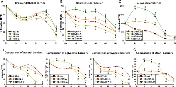
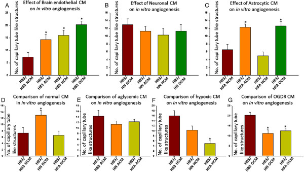
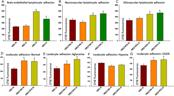
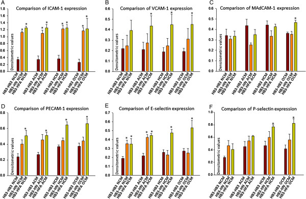
Neurovascular and gliovascular interactions significantly affect endothelial phenotype. Physiologically, brain endothelium attains several of its properties by its intimate association with neurons and astrocytes. However, during cerebrovascular pathologies such as cerebral ischemia, the uncoupling of neurovascular and gliovascular units can result in several phenotypical changes in brain endothelium. The role of neurovascular and gliovascular uncoupling in modulating brain endothelial properties during cerebral ischemia is not clear. Specifically, the roles of metabolic stresses involved in cerebral ischemia, including aglycemia, hypoxia and combined aglycemia and hypoxia (oxygen glucose deprivation and re-oxygenation, OGDR) in modulating neurovascular and gliovascular interactions are not known. The complex intimate interactions in neurovascular and gliovascular units are highly difficult to recapitulate in vitro. However, in the present study, we used a 3D co-culture model of brain endothelium with neurons and astrocytes in vitro reflecting an intimate neurovascular and gliovascular interactions in vivo. While the cellular signaling interactions in neurovascular and gliovascular units in vivo are much more complex than the 3D co-culture models in vitro, we were still able to observe several important phenotypical changes in brain endothelial properties by metabolically stressed neurons and astrocytes including changes in barrier, lymphocyte adhesive properties, endothelial cell adhesion molecule expression and in vitro angiogenic potential.


Figures

Figure 1
Figure 1
Differential effects of metabolic stresses on brain endothelial, neurovascular and gliovascular barriers. A)Brain endothelial barrier. Significant differences were observed between normal and OGDR brain endothelial barrier at 4d. B. Neurovascular barrier. Significant differences between aglycemic, hypoxic and OGDR treated neurovascular barrier was observed from 1d until 5d compared to untreated neurovascular barrier. C. Gliovascular barrier. While no significant differences between untreated and hypoxic gliovascular barrier were observed, aglycemic and OGDR gliovascular barrier was significantly different from untreated from 1d to 3d. D. Comparison of untreated brain endothelial, neurovascular and gliovascular barriers. No significant differences between brain endothelial, neurovascular and gliovascular barriers were observed until 2d. However, neurovascular barrier was significantly lower compared to brain endothelial barrier until 4d and gliovascular barrier was significantly lower until 5d. E. Comparison of aglycemic brain endothelial, neurovascular and gliovascular barriers. No significant differences between aglycemic brain endothelial and neurovascular barriers were observed. However, significant increase in aglycemic gliovascular barrier was observed until 3d. Aglycemic gliovascular barrier was significantly lower at 4d and 5d compared to aglycemic brain endothelial barrier. F. Comparison of Hypoxic brain endothelial, neurovascular and gliovascular barriers. While hypoxic neurovascular barrier was significantly lower than hypoxic brain endothelial barrier from 3d, hypoxic gliovascular barrier was significantly lower from 1d until 5d. G. Comparison of OGDR brain endothelial, neurovascular and gliovascular barriers. Both OGDR neurovascular and gliovascular barriers showed a significant increase compared to OGDR brain endothelial barrier until 3d. However, at 4d OGDR gliovascular barrier was significantly lower compared to OGDR brain endothelial and neurovascular barriers. Repeated measures ANOVA from 0h baseline. Values are expressed in percent baseline at 0h ± SEM. Un-paired t-test was used to check significance between groups at the same time point. # P<0.05 is considered significantly different from respective controls at the same time point.
Figure 2
Figure 2
Differential effects of metabolic stresses on neuronal and astrocytic interactions in modulating brain endothelial angiogenic potential. A. Effect of metabolically stressed brain endothelial CM (self-conditioned medium) on brain endothelial angiogenesis. Significant increase in number of brain endothelial vessel like structures were observed with self-CM obtained from aglycemic, hypoxic and OGDR treated brain endothelial cells compared to untreated brain endothelial CM. B. Effect of metabolically stressed neuronal CM on brain endothelial angiogenesis. No significant difference in number of vessel like structures were observed with neuronal aglycemic, hypoxic or OGDR CM compared to untreated neuronal CM. C. Effect of metabolically stressed astrocytic CM on brain endothelial angiogenesis. Significant increase in number of vessel like structures were observed with astrocytic aglycemic and OGDR CM compared to untreated astrocyte CM. No significant differences were observed between astrocytic hypoxic CM and untreated astrocytic CM. D. Comparison of untreated CM. A significant increase in vessel like structures were observed with untreated neuronal CM compared to untreated brain endothelial CM. No differences between untreated astrocytic CM and untreated brain endothelial CM was observed. E. Comparison of aglycemic CM. No significant differences were observed between aglycemic brain endothelial, neuronal and astrocytic CM. F. Comparison of hypoxic CM. A significant decrease in vessel like structures were observed with hypoxic neuronal and astrocytic CM compared to hypoxic brain endothelial CM. G. Comparison of OGDR CM from brain endothelial cells, neurons and astrocytes. A significant decrease in vessel like structures were observed with OGDR neuronal and astrocytic CM compared to OGDR brain endothelial CM. One way ANOVA with Bonferroni post testing was used to check significance between 2 specific groups. Un-paired t-test was used to check statistical significance between two groups.*P<0.05 is considered significantly different from controls.
Figure 3
Figure 3
Differential effects of metabolic stresses on brain endothelial, neurovascular and gliovascular-leukocyte adhesive interactions. A. Effect of metabolic stresses on brain endothelial-lymphocyte adhesion. Hypoxic and OGDR treated brain endothelial cells significantly promoted lymphocyte adhesion compared to untreated and aglycemic brain endothelial cells. B. Effect of metabolic stresses on neurovascular-lymphocyte adhesion. Similar to brain endothelial-lymphocyte adhesion, hypoxia and OGDR significantly induced neurovascular-lymphocyte adhesion compared to untreated and aglycemic neurovascular-lymphocyte adhesion. C. Effect of metabolic stresses on gliovascular-lymphocyte adhesion. Similar to brain endothelial and neurovascular-lymphocyte adhesion, hypoxia and OGDR significantly induced gliovascular-lymphocyte adhesion compared to untreated and aglycemic gliovascular-lymphocyte adhesion. D. Comparison of untreated brain endothelial, neurovascular and gliovascular-lymphocyte adhesion. A significant increase in neurovascular and gliovascular-lymphocyte adhesion was observed compared to untreated brain endothelial cells. E. Comparison of aglycemic brain endothelial, neurovascular and gliovascular-lymphocyte adhesion. A significant increase in aglycemic neurovascular and gliovascular-lymphocyte adhesion was observed compared to aglycemic brain endothelial cells. F. Comparison of hypoxic brain endothelial, neurovascular and gliovascular-lymphocyte adhesion. No significant difference in hypoxic neurovascular and gliovascular-lymphocyte adhesion compared to hypoxic brain endothelial cells was observed. G. Comparison of OGDR brain endothelial, neurovascular and gliovascular-lymphocyte adhesion. A significant increase in OGDR neurovascular and gliovascular-lymphocyte adhesion was observed compared to OGDR brain endothelial cells. Un-paired t-test is used to check statistical significance between two groups. *P < 0.05 considered significantly different from controls.
Figure 4
Figure 4
Differential brain ECAM expression by metabolically stressed brain endothelial, neuronal and astrocyte secreted factors. A. Comparison of brain endothelial ICAM-1 expression. A significant increase in brain endothelial ICAM-1 expression was observed with CM obtained from astrocytes and neurons from all conditions compared to respective brain endothelial CM. B. Comparison of brain endothelial VCAM-1 expression. While no significant increase in brain endothelial VCAM-1 expression was observed with any of neuronal CM, hypoxic and OGDR astrocytic CM significantly increased brain endothelial VCAM-1 expression compared to respective brain endothelial CM. C. Comparison of brain endothelial MAdCAM-1 expression. No significant difference in brain endothelial MAdCAM-1 expression was observed with CM obtained from astrocytes and neurons from any of conditions compared to respective brain endothelial CM. D. Comparison of brain endothelial PECAM-1 expression. While no significant difference in brain endothelial MAdCAM-1 expression was observed with any of neuronal CM, astrocytic CM from all conditions significantly increased brain endothelial PECAM-1 expression compared to respective brain endothelial CM. E. Comparison of brain endothelial E-selectin expression. A significant increase in brain endothelial E-selectin was observed with both normal and aglycemic neuronal and astrocytic CM compared to respective brain endothelial CM. However, while neither hypoxic nor OGDR neuronal CM increased brain E-selectin expression, both hypoxic and OGDR astrocytic CM significantly increased E-selectin expression compared to respective brain endothelial CM. F. Comparison of brain endothelial P-selectin expression. Except OGDR astrocytic CM, none of the other conditions significantly induced brain endothelial P-selectin expression compared to respective brain endothelial CM. Un-paired t-test was used to check significance between 2 specific groups. *P < 0.05 considered significantly different from controls.

Similar articles

Cited by

References

    1. Kim JH, Kim JH, Park JA, Lee SW, Kim WJ, Yu YS. et al.Blood-neural barrier: intercellular communication at glio-vascular interface. J Biochem Mol Biol. 2006;39:339–345. doi: 10.5483/BMBRep.2006.39.4.339. - DOI - PubMed
    1. Masamoto K, Tomita Y, Toriumi H, Aoki I, Unekawa M, Takuwa H. et al.Repeated longitudinal in vivo imaging of neuro-glio-vascular unit at the peripheral boundary of ischemia in mouse cerebral cortex. Neuroscience. 2012;212:190–200. - PubMed
    1. Stanimirovic DB, Friedman A. Pathophysiology of the neurovascular unit: disease cause or consequence? J Cereb Blood Flow Metab. 2012;32:1207–1221. doi: 10.1038/jcbfm.2012.25. - DOI - PMC - PubMed
    1. Zhang JH, Badaut J, Tang J, Obenaus A, Hartman R, Pearce WJ. The vascular neural network–a new paradigm in stroke pathophysiology. Nat Rev Neurol. 2012;8:711–716. doi: 10.1038/nrneurol.2012.210. - DOI - PMC - PubMed
    1. Pelligrino DA, Vetri F, Xu HL. Purinergic mechanisms in gliovascular coupling. Semin Cell Dev Biol. 2011;22:229–236. doi: 10.1016/j.semcdb.2011.02.010. - DOI - PMC - PubMed

Publication types

MeSH terms

LinkOut - more resources