. 2014 Jan 16;40(1):78-90.
doi: 10.1016/j.immuni.2013.10.023.

Signaling through the adaptor molecule MyD88 in CD4+ T cells is required to overcome suppression by regulatory T cells

Affiliations

Signaling through the adaptor molecule MyD88 in CD4+ T cells is required to overcome suppression by regulatory T cells

Dominik Schenten et al. Immunity. .

Erratum in

  • Immunity. 2014 May 15;40(5):814

Abstract

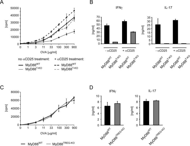
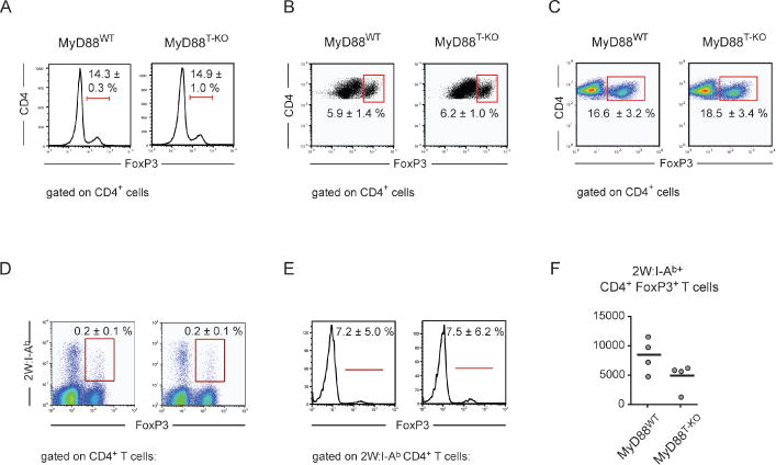
Innate immune recognition controls adaptive immune responses through multiple mechanisms. The MyD88 signaling adaptor operates in many cell types downstream of Toll-like receptors (TLRs) and interleukin-1 (IL-1) receptor family members. Cell-type-specific functions of MyD88 signaling remain poorly characterized. Here, we have shown that the T cell-specific ablation of MyD88 in mice impairs not only T helper 17 (Th17) cell responses, but also Th1 cell responses. MyD88 relayed signals of TLR-induced IL-1, which became dispensable for Th1 cell responses in the absence of T regulatory (Treg) cells. Treg cell-specific ablation of MyD88 had no effect, suggesting that IL-1 acts on naive CD4(+) T cells instead of Treg cells themselves. Together, these findings demonstrate that IL-1 renders naive CD4(+) T cells refractory to Treg cell-mediated suppression in order to allow their differentiation into Th1 cells. In addition, IL-1 was also important for the generation of functional CD4(+) memory T cells.


Figures

Figure 1
Figure 1. Impaired CD4+ T cells response in MyD88T-KO mice
(A) Proliferation of CD4+ T cells from MyD88T-KO mice and wild-type controls. CD4+ T cells were isolated from the draining lymph nodes 7 days after immunization in the feet with OVA + LPS in IFA and re-stimulated with OVA in the presence of irradiated splenocytes. Proliferation was measured 3 days later by 3H-thymidine incorporation. A representative of 3 experiments is shown. (B). Cytokine secretion of CD4+ T cells following restimulation with OVA. (C). TH1 response in MyD88T-KO mice and wild-type controls after immunization with 2W peptide + LPS in IFA and restimulation with 2W peptide. (D). Frequency of 2W:I-Ab+ CD4+ T cells in MyD88T-KO and wild-type control mice T cells 7 days after footpad immunization with 2W peptide + LPS in IFA. (E). Cytokine production of 2W:I-Ab+ CD4+ T cells in MyD88T-KO and wild-type control mice after restimulation with an αCD3e antibody. (F). Absolute numbers of all cells, CD4+ T cells, and 2W:I-Ab+ CD4+ T cells in the draining lymph nodes of MyD88T-KO and wild-type controls immunized with 2W peptide + LPS in IFA 7 days earlier.
Figure 2
Figure 2. Proliferation, viability, and differentation of 2W:IA(b)+ CD4+ T cells from MyD88T-KO and wild-type control mice
(A) Proliferation of antigen-specific 2W:I-A(b)+ CD4+ T cells in the draining lymph nodes of MyD88T-KO and wild-type control mice between day 4–7 after immunization with 2W peptide + LPS in IFA as measured by incorporation of BrdU. Left panel: A representative experiment is shown. Right panel: Statistical representation of all experiments normalized to the level of BrdU incorporation of 2W:I-A(b)+ CD4+ T cells of MyD88WT mice. Each dot represents one mouse. (B) Frequency of apoptotic antigen-specific 2W:I-A(b)+ CD4+ T cells in the draining lymph nodes of MyD88T-KO and wild-type control mice on day 7 after immunization with 2W peptide + LPS in IFA, measured by staining for active caspase-3. Shown is the mean ± standard deviation. (C) T-bet expression by qPCR in immunized CD4+ T cells in the draining lymph nodes of MyD88T-KO and wild-type control mice. A representative experiment using the pooled samples from 5–10 mice per genotype is shown.
Figure 3
Figure 3. Impaired CD4+ T cells response in MyD88T-KO mice is caused by defective IL-1 signaling
(A, B) Expression of the receptors for IL-1 (A) and IL-18 (B) in CD4+ T cells in the draining lymph nodes of immunized MyD88T-KO and wild-type control mice as measured by qPCR. The expression levels were normalized to the level of naïve CD4+ T cells. A representative experiment using the pooled samples from 5–10 mice per genotype is shown. (C, D) CD4+ T cell response of Rag2-deficient mice reconstituted with a 3:1 mix of bone marrow from TCRβ-deficient mice and IL1RKO, IL18KO, or TLR2KO; TLR4KO mice, respectively. CD4+ T cells were restimulated with OVA in the presence of irradiated splenocytes 7 days after the immunzation of the bone marrow chimeras in the feet with OVA + LPS in IFA. The proliferation was measured 3 days later by 3H-thymidine incorporation (C) and the secretion of IFNγ and IL-17 of the restimulated CD4+ T cells was measured by ELISA (D). A epresentative experiment is shown. (E, F). CD4+ T cell response of mixed bone marrow chimeras harboring bone marrow of IL1RKO, IL18KO, or wild-type mice mixed with bone marrow of TCRβKO mice in a ratio of 1:3 after footpad immunization with OVA + LPS in IFA. A representative experiment is shown.
Figure 4
Figure 4. Induction of a TH1 response requires T cell-specific MyD88 signaling in naïve or effector CD4+ T cells in order to overcome Treg-mediated suppression
(A, B) Restoration of the TH1 response after transient depletion of CD25+ Tregs in MyD88T-KO mice. MyD88T-KO and control mice were immunized with OVA + LPS in IFA in the footpads following the injection of an αCD25 antibody 3 days earlier in order to deplete CD25+ Tregs. CD4+ T cells were isolated 7 days after immunization and restimulated with OVA in vitro in the presence of irradiated splenocytes. Proliferation (A) and cytokine secretion (B) of the CD4+ T cells were measured 3 days later. A representative experiment is shown. (C, D) CD4+ T cell response in MyD88TREG-KO mice and wild-type controls. The mice were immunized with OVA + LPS in IFA, the proliferation (C) and cytokine secretion (D) was measured as described before. A representative experiment is shown.
Figure 5
Figure 5. Frequency of Tregs in MyD88T-KO mice
(A) Frequency of FoxP3+ Tregs as percentage of CD4+ T cells in unimmunized wild-type and MyD88T-KO mice. A representative of 3 independent experiments is shown. (B) Frequency of FoxP3+ Tregs after immunization with OVA + LPS in IFA and subsequent restimulation of isolated CD4+ T cells with OVA in the presence of irradiated splenocytes. An experiment representing 3 independent experiments is shown. (C) Frequency of FoxP3+ Tregs in wild-type and MyD88T-KO mice in the draining lymph nodes 7 days after immunization with OVA + LPS in IFA. A representative of three experiments is shown. (D, E) Frequency of antigen-specific FoxP3+ 2W:I-A(b)+ Tregs in the draining lymph nodes 7 days after immunization with 2W peptide + LPS in IFA, shown as percentage of all CD4+ T cells (D) or percentage of all 2W:I-A(b)+ CD4+ T cells (E). A representative of 3 experiments is shown. (F) Absolute numbers of FoxP3+ 2W:I-A(b)+ Tregs in the draining lymph nodes 7 days after immunization with 2W peptide + LPS in IFA.
Figure 6
Figure 6. Memory CD4+ T cell response in MyD88T-KO mice
(A, B) Frequency of 2W:I-A(b)+ memory CD4+ T cells in MyD88T-KO mice and wild-type controls. The frequency of 2W:I-A(b)+ CD4+ T cells and 2W:I-A(b)+ memory CD4+ T cells was determined 40–60 days after immunization with 2W peptide + LPS in IFA. The mice were immunized either in the presence (− αCD25) or transient absence (+ αCD25) of Tregs during the primary immunization. (A). Frequency of 2W:I-A(b)+ CD4+ T cells as a percentage of total CD4+ T cells (upper panels) and frequency of 2W:I-A(b)+ CD44+ CD62L+ memory CD4+ T cells as a percentage of total 2W:I-A(b)+ CD4+ T cells (lower panels). Values represent mean ± standard error. (B). Frequency of 2W:I-A(b)+ CXCR5 PD-1 effector memory CD4+ T, 2W:I-A(b)+ CXCR5+ PD-1 central memory CD4+ T cells, and 2W:I-A(b)+ CXCR5+ PD-1+ T follicular helper (TFH) cells as a percentage of total 2W:I-A(b)+ CD4+ T cells. A representative experiment is shown. (C, D). CD4+ memory T cell response after transient depletion of CD25+ Tregs prior to both the primary and secondary immunization. MyD88T-KO mice and control mice were injected with an αCD25 antibody 3 days prior to the primary immunization with OVA + LPS in IFA. After 60 days, Tregs were transiently depleted again and the mice were challenged 3 days later with OVA + LPS in IFA. CD4+ T cells were isolated 7 days later and restimulated with OVA in vitro in the presence of irradiated splenocytes. The proliferation measured by 3H-thymidine incorporation (C) and cytokine secretion measured by ELISA are shown (D). The data reflect representative results from 2 independent experiments involving a total of 4 mice per genotype.
Figure 7
Figure 7. Gene expression analysis of MyD88T-KO and IL6RαT-KO mice compared to wild-type controls
(A) RNASeq of antigen-specific 2W:I-A(b)+ CD4+ T cells from the draining lymph nodes of MyD88T-KO, IL6RαT-KO, and wild-type control mice that were isolated by flow cytometry 7 days after immunization with 2W peptide + LPS in IFA. (B) Principal component analysis MyD88T-KO, IL6RαT-KO, and wild-type control mice.

Similar articles

Cited by

References

    1. Acosta-Rodriguez EV, Napolitani G, Lanzavecchia A, Sallusto F. Interleukins 1beta and 6 but not transforming growth factor-beta are essential for the differentiation of interleukin 17-producing human T helper cells. Nat Immunol. 2007;8:942–949. - PubMed
    1. Ben Ahmed M, Belhadj Hmida N, Moes N, Buyse S, Abdeladhim M, Louzir H, Cerf-Bensussan N. IL-15 renders conventional lymphocytes resistant to suppressive functions of regulatory T cells through activation of the phosphatidylinositol 3-kinase pathway. J Immunol. 2009;182:6763–6770. - PubMed
    1. Ben-Sasson SZ, Hu-Li J, Quiel J, Cauchetaux S, Ratner M, Shapira I, Dinarello CA, Paul WE. IL-1 acts directly on CD4 T cells to enhance their antigen-driven expansion and differentiation. Proc Natl Acad Sci U S A. 2009;106:7119–7124. - PMC - PubMed
    1. Bettelli E, Carrier Y, Gao W, Korn T, Strom TB, Oukka M, Weiner HL, Kuchroo VK. Reciprocal developmental pathways for the generation of pathogenic effector TH17 and regulatory T cells. Nature. 2006;441:235–238. - PubMed
    1. Brodsky IE, Monack D. NLR-mediated control of inflammasome assembly in the host response against bacterial pathogens. Semin Immunol. 2009;21:199–207. - PubMed

Publication types

MeSH terms