. 2014 Jan 16;40(1):91-104.
doi: 10.1016/j.immuni.2013.11.019.

Embryonic and adult-derived resident cardiac macrophages are maintained through distinct mechanisms at steady state and during inflammation

Affiliations

Embryonic and adult-derived resident cardiac macrophages are maintained through distinct mechanisms at steady state and during inflammation

Slava Epelman et al. Immunity. .

Abstract

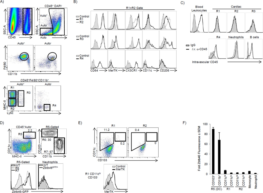
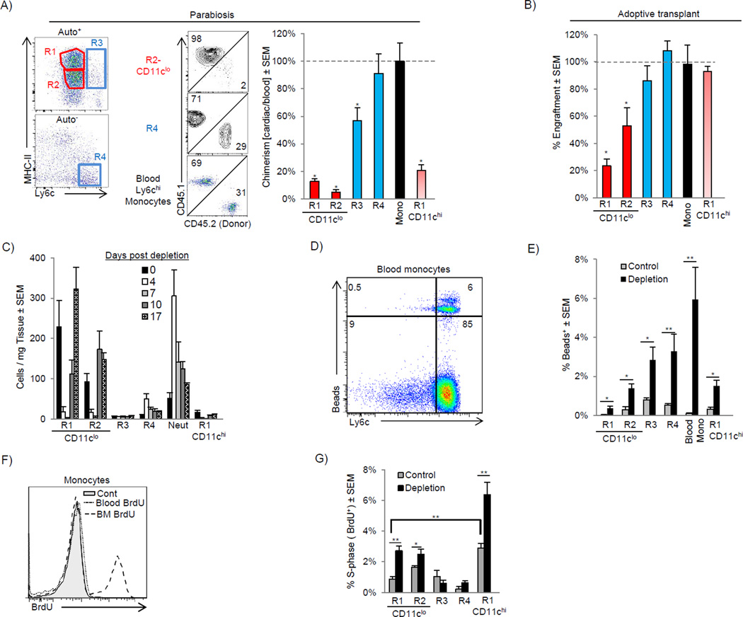
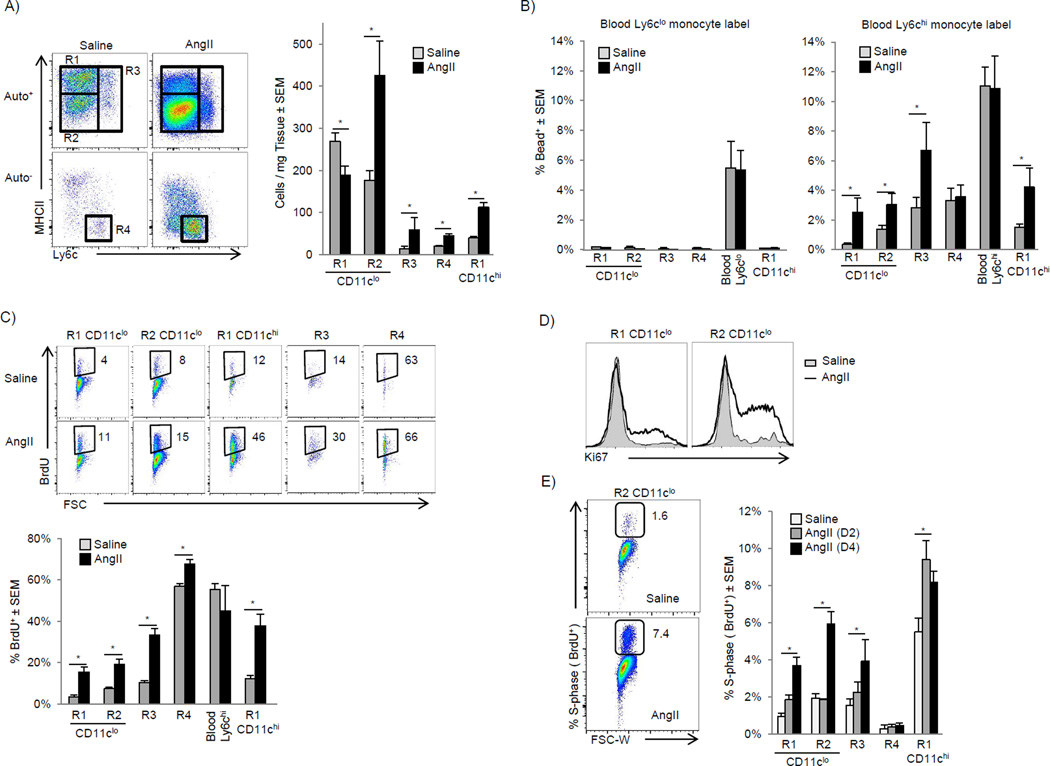
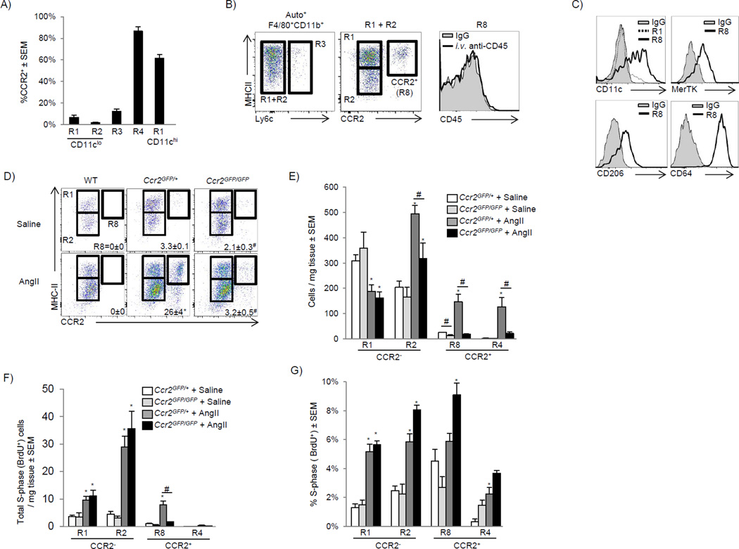
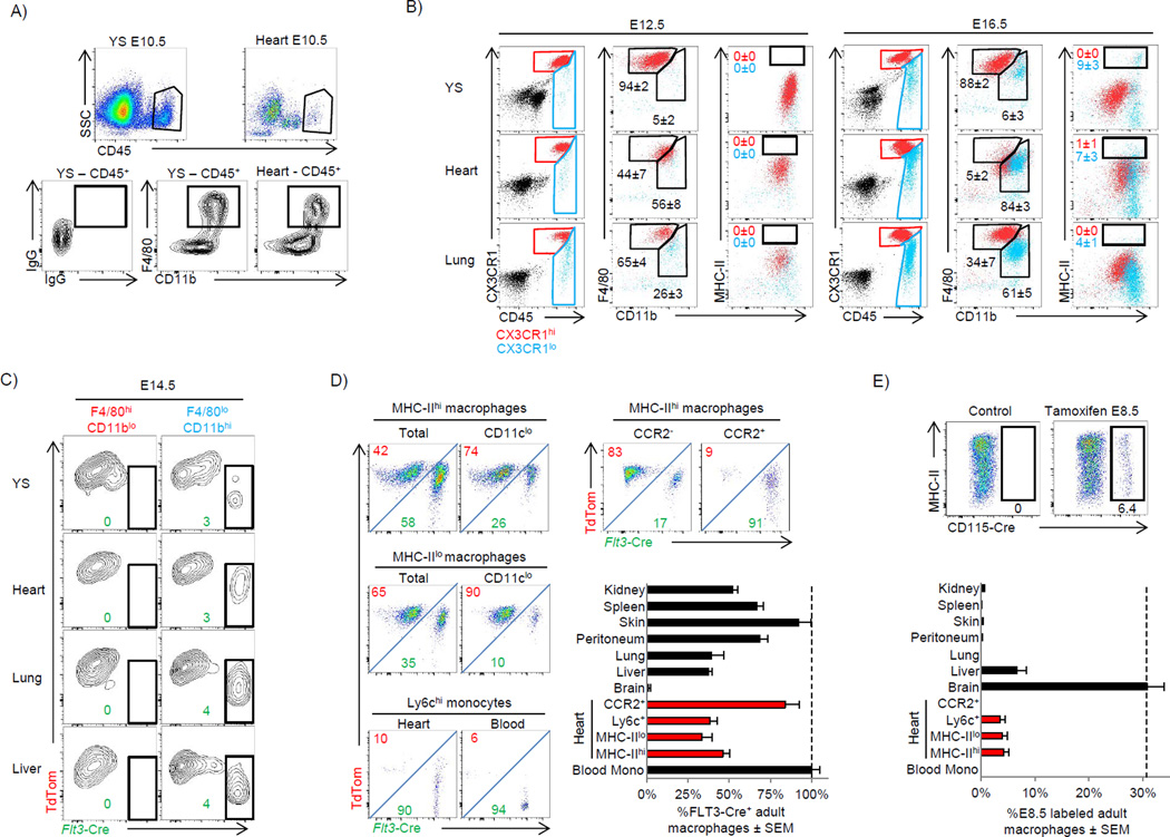
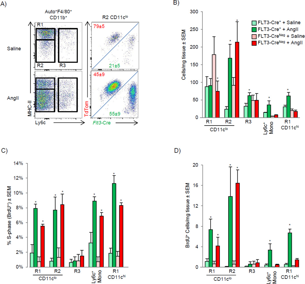
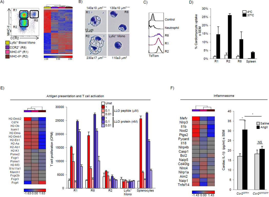
Cardiac macrophages are crucial for tissue repair after cardiac injury but are not well characterized. Here we identify four populations of cardiac macrophages. At steady state, resident macrophages were primarily maintained through local proliferation. However, after macrophage depletion or during cardiac inflammation, Ly6c(hi) monocytes contributed to all four macrophage populations, whereas resident macrophages also expanded numerically through proliferation. Genetic fate mapping revealed that yolk-sac and fetal monocyte progenitors gave rise to the majority of cardiac macrophages, and the heart was among a minority of organs in which substantial numbers of yolk-sac macrophages persisted in adulthood. CCR2 expression and dependence distinguished cardiac macrophages of adult monocyte versus embryonic origin. Transcriptional and functional data revealed that monocyte-derived macrophages coordinate cardiac inflammation, while playing redundant but lesser roles in antigen sampling and efferocytosis. These data highlight the presence of multiple cardiac macrophage subsets, with different functions, origins, and strategies to regulate compartment size.


Figures

Figure 1
Figure 1. The adult heart contains distinct cardiac macrophage subsets (see also Fig S1)
Cardiac single cell suspensions were analyzed by flow cytometry – see full gating strategy in Fig S1A. A) CD45+ leukocytes were identified, doublets excluded (by FSC-W vs. FSC-A) and dead cells excluded by DAPI. Live cells were stratified by autofluorescence (Auto+ or Auto), gated on F4/80+ CD11b+ myeloid cells and further stratified by MHC-II and Ly6c expression. R1: MHC-IIhi macrophages. R2: MHC-IIlo macrophages. R3: Ly6c+ macrophages. R4: Ly6chi monocytes. B) Cardiac samples were labeled with isotype control antibody (Control) or with the indicated antibodies. Expression of CX3CR1 was assessed in Cx3cr1GFP/+ mice and compared with WT mice (Control). C) To label intravascular leukocytes, mice were injected i.v. with anti-CD45 and sacrificed 5 min later. Cells were gated as in panel A and intravascular CD45 fluorescence is shown. B cells were B220+ MHCII+ CD11bF4/80 and neutrophils were Ly6g+ CD11b+ F4/80. D) The Auto subset contained the majority of cardiac DCs (Total DCs - R5), which were made up of CD103+ CD11b (R6) and CD103CD11bHi (R7) DCs. ZBTB46 expression was assessed in Zbtb46GFP/+ mice in R6 and R7, and compared to Ly6g+ neutrophils and compared to WT mice. E) Expression of CD11c and CD103 within the primary macrophage gates (R1 and R2). F) Relative Zbtb46 fluorescence ratio in myeloid subsets within the myocardium. The geometric mean fluorescence intensity (gMFI) in each subset in Zbtb46GFP/+ mice was divided by gMFI of that subset in WT mice. The primary macrophages populations in R1 and R2 were further stratified by CD11c expression. N=4–8.
Figure 2
Figure 2. Distinct mechanisms regulate cardiac macrophage turnover in steady and following the disruption of homeostasis
A) CD45.1 and CD45.2 mice were surgically joined to create parabiotic mice and were analyzed after 2 weeks. Dot plots show rate of chimerism for representative populations. The percentage chimerism for each cardiac monocyte and macrophage subset was normalized for the chimeric rate for blood monocytes and expressed as a percentage. B) E14.5 Fetal liver CD150+ HSCs were sorted and adoptively transplanted into sublethally irradiated mice. 16 weeks after transplant, mice were harvested and engraftment of transplanted cells was assessed in blood monocytes and cardiac monocytes and macrophage populations. Engraftment for cardiac populations was normalized for blood monocyte engraftment. C–G) Mice were injected with either control liposomes (Control) or liposomes containing clodronate to deplete cardiac macrophages (Depletion) and analyzed over time (C). D–E) Ly6cHi blood monocytes were labeled with fluorescent microspheres in vivo after macrophage depletion (Tacke et al., 2006). D) Bead expression in SSCLowCD115+F4/80+ blood monocytes after macrophage depletion. E) Percentage of cardiac bead+ cells after depletion. F) To assess proliferation, mice were injected with BrdU and analyzed 2 hrs post injection. Proliferation (BrdU+) in Ly6chi monocytes in the bone marrow (BM) and blood is shown. G) Percentage of proliferating BrdU+ cells following macrophage depletion (7 days) within the myocardium in each subset. N=4–7, * P <0.05. ** P<0.01. Data represents at least two experiments, n= 6–12 mice per group.
Figure 3
Figure 3. AngII infusion induces numerical expansion of cardiac macrophage through monocytes recruitment and local proliferation (see also Fig S2)
A–E). WT mice were implanted with pumps containing either saline or AngII (2 mg/kg/day). A) Flow cytometric profiles 4 days post infusion gated as in Fig 1A, and graphed numerically. B) Mice with in vivo bead-labeled Ly6clo or Ly6chi monocytes were implanted with pumps as above, blood and cardiac tissue was analyzed on day 4 and the percentage of bead+ cells is shown. C) Osmotic pumps containing either saline or AngII (2 mg/kg/day) were implanted and Ly6chi monocytes were labeled by injecting mice with BrdU 48 and 24 hrs prior to harvest. Mice were sacrificed 3 days after pump implantation and BrdU detected by flow cytometry. D) Intracellular Ki-67 was detected by flow cytometry 4 days after pump implantation as Fig 3A. E) Percentage of proliferating cells in s-phase was assessed by a single BrdU pulse 2 hrs prior to harvest. Mice were sacrificed 2 or 4 days after pump implantation. Representative flow cytometric profiles at day 4 in MHC-IIlo CD11clo macrophages (R2) and the percentage of BrdU+ cells in each gate. * P<0.05. 2–4 independent experiments, 4–7 mice per group.
Figure 4
Figure 4. CCR2 deficient mice distinguish local macrophage expansion from influx of peripheral monocytes (see also Fig S3)
A) Cardiac single cell suspensions from Ccr2GFP/+mice were gated as in Fig 1A and percentage of CCR2+ cells in each gate is shown. B) Ly6c MHC-IIhi and MHC-IIlo macrophages were gated together (R1+R2) in order identify CCR2+ macrophages (R8). To determine if CCR2+ macrophages were extravascular, mice were injected with anti-CD45 i.v. as in Fig 1C and intravascular CD45 expression is shown. C) Expression of CD11c, MerTK, CD206 and CD64 was assessed in the regions as outlined. D–G) Either saline or AngII (1.5 mg/kg/day) containing pumps were implanted and cardiac tissue analyzed at day 4 in either Ccr2GFP/+ or Ccr2GFP/GFP mice. D) Cells were gated as in Fig 4B and the percentage of CCR2+ macrophages (R8) is given. E) Total cell numbers, including gating on cardiac Auto Ly6c+CCR2+ monocytes (R4). F) Total number of proliferating cells (BrdU+, S-phase) per mg of tissue after a 2 hr BrdU pulse, and G) the percentage of cells in S-phase in each subset. 2–4 independent experiments, 4–8 mice per group. * P<0.05 vs. saline, # P<0.05 vs. Ccr2GFP/+.
Figure 5
Figure 5. Prenatal macrophages colonize the embryonic heart and persist into adulthood (see also Fig S4 and S5)
A–B) Cx3cr1GFP/+ embryos were extracted from pregnant females at either E10.5, E12.5 or E16.5. A) Comparison of macrophage populations between the yolk sac (YS) and heart tissue at E10.5. Red color indicates CD45+CX3CR1hi and blue indicates CD45+CX3CR1lo cells. B) Comparison of yolk sac, heart and lung macrophages at E12.5 and E16.5. C) Flt3-Cre Rosa-mTmG mice were analyzed for reporter expression (E14.5) in embryonic macrophages from various tissues. 2–4 litters for each time point with pooled tissue from 2–6 embryos per animal were used. D) Flow plots showing of adult cardiac macrophages populations in Flt3-Cre Rosa-mTmG mice. Graphed data represent FLT3-derived cardiac macrophages subsets (red), and other tissue macrophages. Data was normalized to blood monocyte FLT3 recombination, which was typically ∼90%. MHC-IIHi and MHC-IILow macrophages were gated on the CD11clo subsets as in Fig 2A. CCR2+ macrophages (R8) were gated as in Fig 4B. E) CD115-Mer-iCre-Mer Rosa-mTmG mice were gavaged with tamoxifen at E8.5 to label the progeny of yolk sac macrophages. Mice were sacrificed at 10 weeks of age to determine whether yolk sac macrophage progeny persisted into adulthood. Cardiac macrophages were gated on CD45+ Auto+ F4/80+ CD11b+ Ly6c CD11clo resident macrophage subsets. Graphed data represent yolk sac-derived cardiac macrophage subsets (red), and other tissue macrophages. See supplemental methods for exact gating strategies for each tissue macrophage population. Experiments were repeated at least twice, n=4–8 animals were group.
Figure 6
Figure 6. Genetic lineage tracing during AngII induced inflammation
A–D) FLT3-Cre x Rosa mTmG mice were analyzed for reporter expression in the myocardium and blood in mice implanted with either saline or AngII containing pumps for 4 days (2 mg/kg/day). A) Representative flow cytometric plots from MHC-IIloCD11clo macrophages and cumulative total cells counts per mg of tissue (B) in each lineage (FLT3-Cre+: Green, FLT3-Cre: Red). C–D) Flt3-Cre x Rosa mTmG mice were pulsed with BrdU 2 hrs prior to harvest. C) Percentage of cells in S-phase, and D) Total number of cells in S-phase in each subset is shown. Experiments were repeated at least twice, n=4–6 animals were group. * P<0.05 vs. saline.
Figure 7
Figure 7. Adult-derived macrophages coordinate cardiac inflammation, while playing redundant but lesser roles in antigen sampling and efferocytosis (see also Fig S6, Table S2 and S3)
Blood Ly6chi monocytes (yellow), MHC-IIhi CCR2 macrophages (R1, Pink) MHC-IIlo CCR2 macrophage (R2, brown) and CCR2+ MHC-IIhi macrophages (R8, purple) were sorted from Ccr2GFP/+ mice (4 replicates), RNA was extracted and global transcriptional profiling performed. A) Hierarchal clustering of 4557 genes differentially expressed in the entire cell population (Fold change >2). B) Sorted macrophage and Ly6chi blood monocyte subsets were stained with Hema3 solution, surface area was calculated based on an average of at least 20 cells, ** P<0.01 vs. Ly6c+ monocytes. C) Expression of TdTom was determined in cardiac macrophages and neutrophils (Ly6g+CD11b+F4/80) from cardiomyocyte restricted reporter mice (Mlc2V-Cre x Rosa-TdTom). Control represents background cardiac macrophage fluorescence from WT mice. D) Fluorescently labeled apoptotic / necrotic cardiomyocytes from WT mice were incubated with cardiac (or splenic) single cell suspension from Ccr2GFP/+ mice for 4 hours, at either 4°C or 37°C to assess phagocytic uptake. The percentage of macrophage that took up labeled cardiomyocytes was determined by flow cytometry. E) Hierarchical clustering of genes regulating antigen processing and presentation in cardiac macrophages. Sorted cardiac macrophages (as in Fig 7A) were incubated with either Listeriolysin O (LLO) peptide (190–201), or LLO protein (WW non-hemolytic variant), and T cell activation was assessed by IL-2 driven H3 thymidine uptake and expressed in counts per minute (see experimental procedures). F) Hierarchical clustering of genes regulating inflammasome activation in cardiac macrophages. In vivo cardiac IL-1β production was measured in cardiac tissue lysate from mice (Ccr2GFP/+ or Ccr2GFP/GFP) infused with either saline or AngII (2 mg/kg/day) for 4 days. Each experiment was repeated at least twice, with 2–6 mice / group. * P<0.05.

Comment in

Similar articles

Cited by

References

    1. Boyer SW, Schroeder AV, Smith-Berdan S, Forsberg EC. All hematopoietic cells develop from hematopoietic stem cells through Flk2/Flt3-positive progenitor cells. Cell Stem Cell. 2011;9:64–73. - PMC - PubMed
    1. Carrero JA, Vivanco-Cid H, Unanue ER. Listeriolysin o is strongly immunogenic independently of its cytotoxic activity. PLoS. One. 2012;7:e32310. - PMC - PubMed
    1. Davies LC, Rosas M, Jenkins SJ, Liao CT, Scurr MJ, Brombacher F, Fraser DJ, Allen JE, Jones SA, Taylor PR. Distinct bone marrow-derived and tissue-resident macrophage lineages proliferate at key stages during inflammation. Nat. Commun. 2013;4:1886. - PMC - PubMed
    1. Francis GS. Neurohormonal control of heart failure. Cleve. Clin. J. Med. 2011;78(Suppl):S75–S79. - PubMed
    1. Frangogiannis NG, Dewald O, Xia Y, Ren G, Haudek S, Leucker T, Kraemer D, Taffet G, Rollins BJ, Entman ML. Critical role of monocyte chemoattractant protein-1/CC chemokine ligand 2 in the pathogenesis of ischemic cardiomyopathy. Circulation. 2007;115:584–592. - PubMed

Publication types

MeSH terms

Associated data