. 2014 Jan 16;156(1-2):146-57.
doi: 10.1016/j.cell.2013.12.017.

Inefficient SRP interaction with a nascent chain triggers a mRNA quality control pathway

Affiliations

Inefficient SRP interaction with a nascent chain triggers a mRNA quality control pathway

Andrey L Karamyshev et al. Cell. .

Abstract

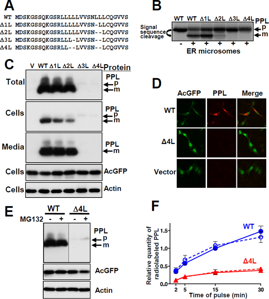
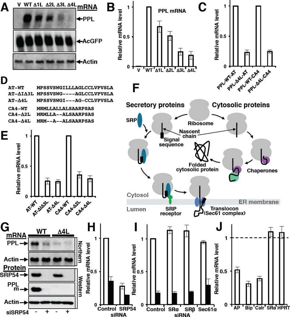
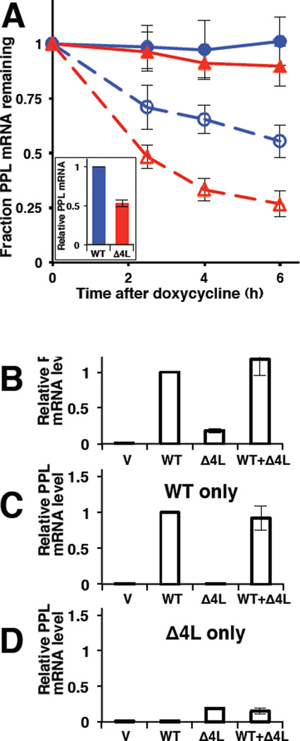
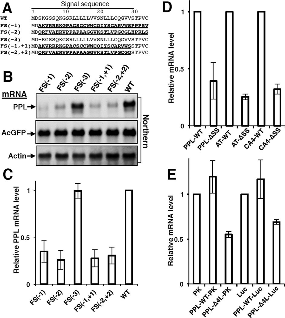
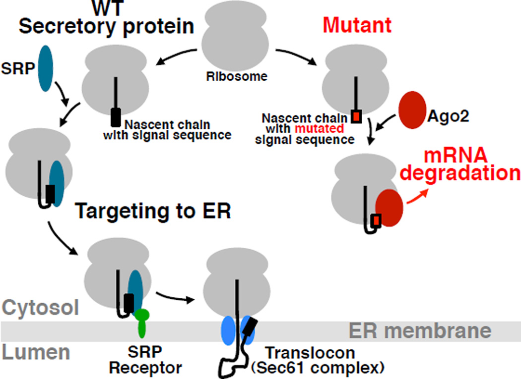
Misfolded proteins are often cytotoxic, unless cellular systems prevent their accumulation. Data presented here uncover a mechanism by which defects in secretory proteins lead to a dramatic reduction in their mRNAs and protein expression. When mutant signal sequences fail to bind to the signal recognition particle (SRP) at the ribosome exit site, the nascent chain instead contacts Argonaute2 (Ago2), and the mutant mRNAs are specifically degraded. Severity of signal sequence mutations correlated with increased proximity of Ago2 to nascent chain and mRNA degradation. Ago2 knockdown inhibited degradation of the mutant mRNA, while overexpression of Ago2 or knockdown of SRP54 promoted degradation of secretory protein mRNA. The results reveal a previously unappreciated general mechanism of translational quality control, in which specific mRNA degradation preemptively regulates aberrant protein production (RAPP).


Figures

Figure 1
Figure 1. Deletions in the PPL Signal Sequence Lead to Defects in Protein Transport and Expression
(A) Signal sequences of WT and mutated PPLs, deletions indicated by dashes. (B) WT and mutant PPLs were synthesized in a rabbit reticulocyte translation system in vitro in the presence or absence of ER microsomes, analyzed by SDS-PAGE, and detected by autoradiography. Positions of mature PPL (m) and precursor (p) are shown. (C and D) WT and mutant PPLs were transiently expressed in HeLa Tet-On cells (cells with empty vector (V) were controls) and detected by Western blot or by immunofluorescence, respectively. AcGFP, expressed from the same plasmids, and endogenous actin were controls. (E) Effect of the proteasome inhibitor MG132 on the level of WT and Δ4L PPLs (detection by Western blot). (F) Pulse-labeling analysis of translated WT (blue circles) and Δ4L (red triangles) PPLs in the presence (dashed lines, n=4) and absence (solid lines, n=5) of MG132 (mean ± SEM). See also Figure S1.
Figure 2
Figure 2. Defects in the Signal Sequence or in a Targeting Factor, SRP, Lead to Decreased Levels of a Secretory Protein mRNA
(A and B) Effect of deletions in signal sequence on PPL mRNA levels. PPL and control mRNAs were analyzed by Northern blot (A) or qPCR (B; n=9, mean ± SD) 42 h after transfection of HeLa Tet-On with WT and mutated PPL plasmids. (C) Presence of a mutated PPL signal sequence (PPL- Δ4L) in hybrid proteins containing the mature part of a1-antitrypsin (AT) or carbonic anhydrase IV (CA4) is sufficient to trigger mRNA depletion. mRNA levels measured by qPCR (n=3, mean ± SD) are shown relatively to mRNA levels of corresponding hybrid proteins containing WT PPL signal sequence (PPL-WT). (D) Deletions (indicated by dashes) in the natural signal sequences of secretory proteins AT or CA4. (E) Deletions in the hydrophobic core of natural signal sequences of AT or CA4 proteins lead to a decrease in their mRNA levels. Graph shows mRNA levels measured by qPCR (n=3, mean ± SD). (F) Scheme shows differences in biogenesis of secretory and cytosolic proteins. When the signal sequence of a secretory protein emerges from the ribosomal tunnel it is recognized by SRP, and the complex is targeted to SRP receptor and finally to a translocon in ER membrane. Nascent chains of cytosolic proteins do not have signal sequences, however, their nascent chains are recognized by ribosome-associated chaperones and that help them fold in the cytosol. (G–H) SRP depletion causes a reduction in the level of secretory protein mRNA. Detection of PPL, and actin mRNAs (Northern blot), SRP54, PPL and actin proteins (Western blot) in HeLa Tet-On cells transfected with siRNA for SRP54 and WT or Δ4L PPL plasmids as indicated (G). Quantification of WT PPL (open bars) or Δ4L mutant (black bars) mRNAs by qPCR in independent sets of SRP54 knockdown experiments (n=3, mean ± SD) (H). (I) Knockdown of SRP receptor subunits SRα and SRβ, or translocon component, Sec61α, does not affect PPL mRNA level (qPCR, n=3, mean ± SD). WT PPL (open bars), Δ4L mutant (black bars). (J) SRP depletion causes reduction in endogenous mRNA levels of secretory and ER proteins. Measured by qPCR (n=3, mean ± SD) mRNA levels in SRP depleted cells are shown relatively those treated with control siRNA (taken as 1). Intestinal alkaline phosphatase (AP) is a secretory protein, Bip and calreticulin (Calr) are ER lumen proteins. mRNA levels of the cytosolic protein HPRT and the SRP-independent ER membrane-associated protein SRα were not altered. See also Figures S2 and Table S1.
Figure 3
Figure 3. Preferential Degradation of Mutant PPL mRNA
(A) qPCR analysis of WT (blue circles) or Δ4L (red triangles) PPL mRNAs under conditions of transcription inhibition (doxycycline treatment, dashed lines and open symbols) or mock (solid lines and symbols). The quantity of mRNA at each time point is shown relative to the initial quantity of the respective mRNA species. Doxycycline was added 20 h after transfection to ensure that adequate levels of Δ4L mRNA still remained at the start of the experiment; the initial WT and Δ4L mutant mRNA content was about 2:1 (inset). The quantity of mRNA at each time point is shown relative to the initial quantity of the respective mRNA species. Data are from 3 experiments, where each time point contained 3 repeats in each experiment (mean ± SD). (B to D) qPCR analysis (n=5, mean ± SD) of PPL WT and Δ4L mRNAs using primers amplifying both WT and mutant (B), only WT (C), or only Δ4L (D) in the cultured human cells expressing WT PPL only (WT), Δ4L mutant only (Δ4L), both WT and Δ4L (WT+ Δ4L), or transfected with empty vector (V). mRNA levels are shown relative to WT, except in D, where mRNA levels were scaled to equate the Δ4L level in D to that in B. See also Figure S3.
Figure 4
Figure 4. Functional Signal Sequence is Required for Protection of the Secretory Protein mRNA from Degradation
(A) N-terminal sequences of PPL mutants with amino acid substitutions in bold and underlined, and deletions as dashes. FS(−1), FS(−2), FS(−3) contain deletions of one, two, three nucleotides, correspondingly, in the third codon. FS(−1, +1), FS(−2,+2) contain additional insertion of one or two nucleotides in the codon 29 to restore original reading frame. mRNAs were analyzed by Northern blots (B) or by qPCR (C; n=3, mean ± SD). (D) Deletions of natural signal sequences from PPL, AT, and CA4 lead to decrease in their mRNA levels (qPCR, n=3, mean ± SD). (E) Presence of a mutated signal sequence in hybrid cytosolic proteins leads to their mRNA depletion (qPCR, n=3, mean ± SD).
Figure 5
Figure 5. Ago2 is in Close Proximity to Mutant Nascent Chains and Associated with Mutant mRNA
(A) Deletions in the PPL signal sequence lead to reduced interaction with SRP54 and increased proximity to a ~100 kDa protein. Photocrosslinking patterns of WT and mutated PPLs are shown. RNCs containing the first 86 residues of WT and mutant PPLs with a photoreactive probe in the signal sequence were produced in vitro in the presence of SRP. Following UV irradiation, samples were analyzed by electrophoresis and autoradiography. The positions of photoadducts containing ~50 kDa (circle) or ~100 kDa (diamond) proteins are shown. (B) Immunoprecipitation (IP) using SRP54 specific antibody demonstrates that the ~50 kDa protein is SRP54. (C) WT PPL crosslinks to the ~100 kDa protein are maximized when lysate is not supplemented with SRP. (D) Scheme of the method for identification of Proteins Interacting with Nascent Chains (iPINCH). (E) Identification of fractions that contain the ~100 kDa protein. Δ4L RNCs were prepared as above and treated with a high salt buffer. The latter were combined with lysate or aliquots of fractionated rabbit reticulocyte ribosome-associated proteins, incubated, UV irradiated and analyzed as in A. The photocrosslinking pattern of the fraction examined by mass spectrometry is boxed. (F) Recombinant Ago2 is in close proximity to mutant nascent chain. Purified recombinant human Ago2 or buffer was added to salt-washed RNCs of Δ4L PPL mutant with photocrosslinking probe. Samples were photocrosslinked and analyzed as above. (G) mRNA of Δ4L PPL is preferentially enriched in Ago2 immunoprecipitates. A short incubation time (20–24 h after plasmid transfection) was used to ensure that significant levels of Δ4L mRNA still remained at the start of the experiment: the WT and Δ4L mutant mRNA content in cell lysates was about 2:1 before IP (inset). Mouse monoclonal Ago2 antibody was used for IP. Data are from two qPCRs, reactions were in triplicates, mean ± SD. See also Figures S4 and Table S2.
Figure 6
Figure 6. Process of mRNA degradation involves Ago2
Ago2 knockdown inhibited reduction of PPL mRNA (A), while overproduction of recombinant WT or mutant D597A Myc-Ago2 reduced the level of PPL mRNA (B). Bars are PPL mRNA levels from Northern blot (n=3 (but n=2 for D597A), mean ± SEM). WT PPL mRNA levels are open bars, Δ4L mutants are black bars. Depletion or overexpression of Ago2 was confirmed by Western blot, actin was a control. See also Figure S5.
Figure 7
Figure 7. Model for Regulation of Aberrant Protein Production (RAPP)
When the nascent chain of a WT secretory protein containing a signal sequence emerges from the ribosome polypeptide exit tunnel during translation it is recognized by SRP. This interaction leads to targeting of the complex to SRP receptor in the ER membrane and finally to the translocon. The nascent chain is co-translationally translocated into the ER lumen, where it is folded with the help of ER chaperones. However, when the nascent chain of a secretory protein contains critical mutations preventing SRP recognition, the unbound nascent chain is exposed outside the polypeptide tunnel. Despite the absence of its interaction with SRP, a defective secretory protein does not typically behave as a cytosolic protein because of the absence of appropriate sequences/conformations/signals required for normal cytosolic nascent chains interactions. Thus, the space normally occupied by SRP at the ribosome remains unoccupied. Ago2 occupies the space and directs mRNA for degradation initiating the RAPP process.

References

    1. Albanese V, Yam AY, Baughman J, Parnot C, Frydman J. Systems analyses reveal two chaperone networks with distinct functions in eukaryotic cells. Cell. 2006;124:75–88. - PubMed
    1. Alder NN, Johnson AE. Cotranslational membrane protein biogenesis at the endoplasmic reticulum. J. Biol. Chem. 2004;279:22787–22790. - PubMed
    1. Ameyar-Zazoua M, Rachez C, Souidi M, Robin P, Fritsch L, Young R, Morozova N, Fenouil R, Descostes N, Andrau JC, et al. Argonaute proteins couple chromatin silencing to alternative splicing. Nat. Struct. Mol. Biol. 2012;19:998–1004. - PubMed
    1. Andrews DW, Lauffer L, Walter P, Lingappa VR. Evidence for a two-step mechanism involved in assembly of functional signal recognition particle receptor. J. Cell Biol. 1989;108:797–810. - PMC - PubMed
    1. Brodsky JL. Cleaning up: ER-associated degradation to the rescue. Cell. 2012;151:1163–1167. - PMC - PubMed

Publication types