. 2014 Jan 16;156(1-2):170-82.
doi: 10.1016/j.cell.2013.11.047.

Potentiated Hsp104 variants antagonize diverse proteotoxic misfolding events

Affiliations

Potentiated Hsp104 variants antagonize diverse proteotoxic misfolding events

Meredith E Jackrel et al. Cell. .

Abstract

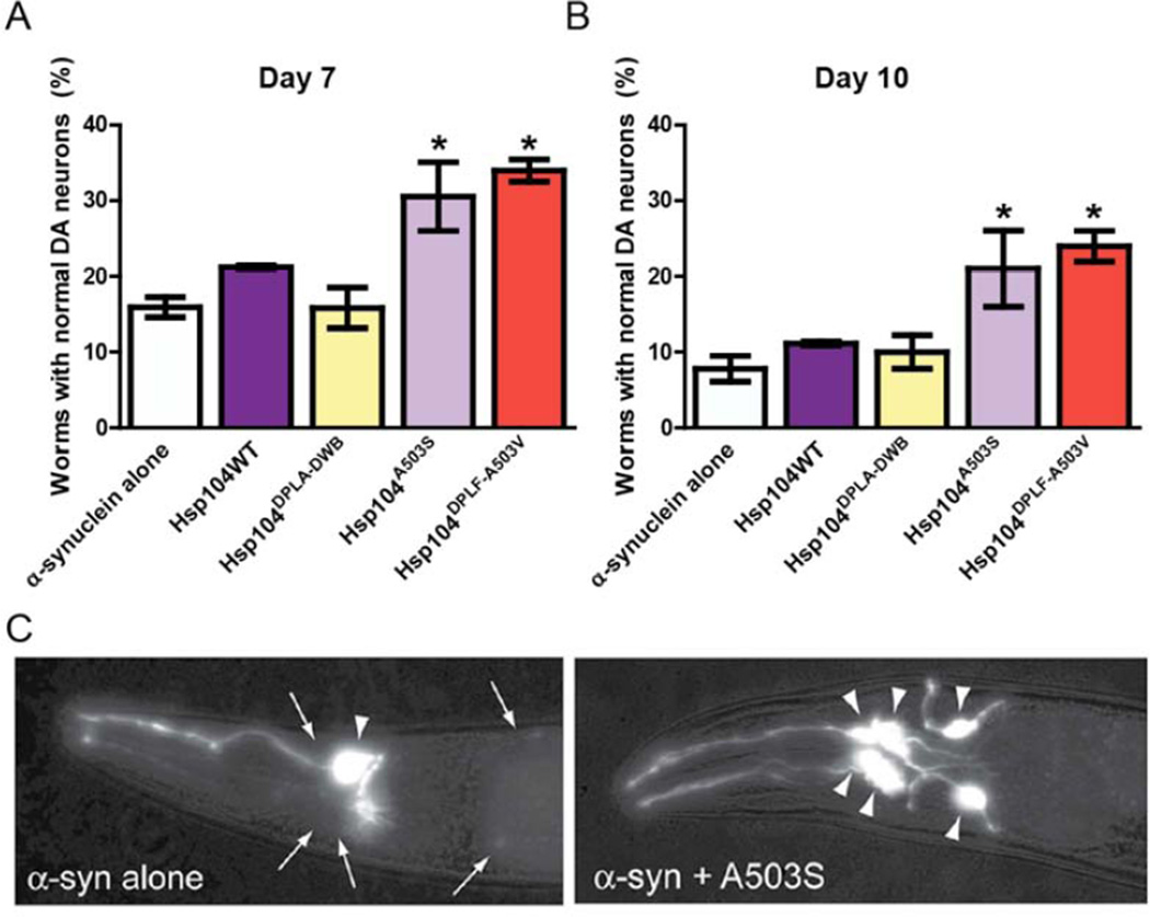
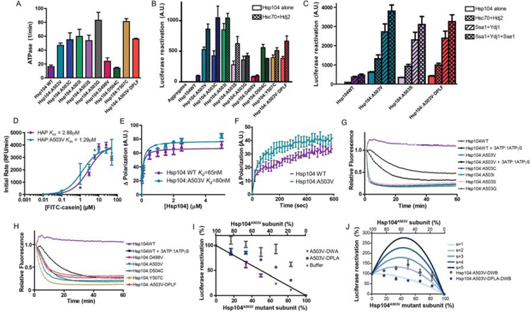
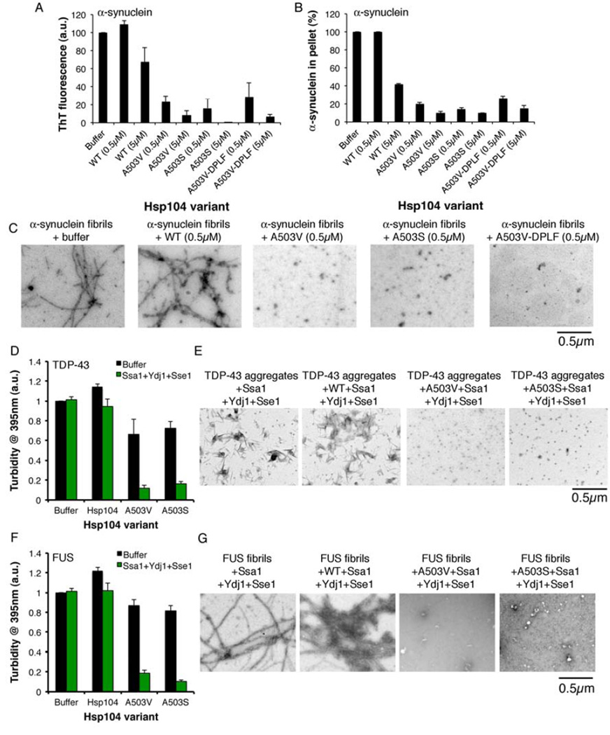
There are no therapies that reverse the proteotoxic misfolding events that underpin fatal neurodegenerative diseases, including amyotrophic lateral sclerosis (ALS) and Parkinson's disease (PD). Hsp104, a conserved hexameric AAA+ protein from yeast, solubilizes disordered aggregates and amyloid but has no metazoan homolog and only limited activity against human neurodegenerative disease proteins. Here, we reprogram Hsp104 to rescue TDP-43, FUS, and α-synuclein proteotoxicity by mutating single residues in helix 1, 2, or 3 of the middle domain or the small domain of nucleotide-binding domain 1. Potentiated Hsp104 variants enhance aggregate dissolution, restore proper protein localization, suppress proteotoxicity, and in a C. elegans PD model attenuate dopaminergic neurodegeneration. Potentiating mutations reconfigure how Hsp104 subunits collaborate, desensitize Hsp104 to inhibition, obviate any requirement for Hsp70, and enhance ATPase, translocation, and unfoldase activity. Our work establishes that disease-associated aggregates and amyloid are tractable targets and that enhanced disaggregases can restore proteostasis and mitigate neurodegeneration.


Figures

Figure 1
Figure 1. Hsp104 MD variants rescue diverse proteotoxicity models
(A) Homology model of the MD and a portion of the small domain of NBD1 of Hsp104. Side chains of key residues are shown as sticks. (B) Δhsp104 yeast strains integrated with galactose-inducible α-syn, FUS, or TDP-43 were transformed with the indicated Hsp104 variant or vector control. Strains were serially diluted fivefold and spotted on glucose (off) or galactose (on) media. (C) Hsp104 variants harboring missense mutations to valine ranging from residue D498 to Y507 were expressed with FUS, TDP-43, or α-syn. (D) Close-up of MD helix 3 from (A). Mutation of D498, A503, D504, or Y507 activates Hsp104. See also Fig. S1.
Figure 2
Figure 2. Hsp104A503X variants suppress TDP-43 toxicity, aggregation, and mislocalization
(A) Δhsp104 yeast transformed with TDP-43 and Hsp104 variants, or YFP and vector, were serially diluted fivefold and spotted onto glucose (off) or galactose (on). (B) Selected strains from (A) were induced in liquid and growth was monitored by A600nm. (C) Strains from (B) were induced for 5h, lysed, and immunoblotted. Uninduced (untreated) and heat shocked cells (HS) serve as controls. 3-Phosphoglycerate kinase (PGK1) serves as a loading control. (D) WT, Δire1, or Δatg8 yeast were co-transformed with vector control, or TDP-43 plus vector, or the indicated Hsp104 variant and were serially diluted fivefold and spotted onto glucose (off) or galactose (on). (E) Fluorescence microscopy of cells co-expressing fluorescently tagged TDP-43 and Hsp104WT, Hsp104A503V, or vector. Cells were stained with DAPI to visualize nuclei (blue). TDP-43 localization was quantified by counting the number of cells containing colocalized nuclear staining. Values represent means±SEM (n=3). (F) Δhsp104 yeast co-transformed with TDP-43 and vector or the indicated Hsp104 variant were induced with galactose for 5h at 30°C, lysed and processed for s edimentation analysis and quantitative immunoblot. The relative amount of insoluble TDP-43 was determined as a % of the vector control. Values represent means±SEM (n=2). See also Fig. S2, S3, and S4.
Figure 3
Figure 3. Hsp104A503X variants suppress FUS toxicity and aggregation
(A) Δhsp104 yeast transformed with FUS and the Hsp104 variants, or YFP and vector, were serially diluted fivefold and spotted onto glucose (off) or galactose (on). (B) Selected strains from (A) were induced in liquid and growth was monitored by A600nm. (C) Strains from (B) were induced for 5h, lysed, and immunoblotted. (D) WT, Δire1, or Δatg8 yeast were co-transformed with vector control, or FUS plus vector, or the indicated Hsp104 variant and were serially diluted fivefold and spotted onto glucose (off) or galactose (on). (E) Fluorescence microscopy of cells co-expressing FUS-GFP and Hsp104WT, Hsp104A503V, or vector. Cells were stained with Hoechst dye to visualize nuclei (blue). FUS aggregation was quantified by counting the number of cells containing 0, 1, or more than 1 foci. Values represent means±SEM (n=3). (F) Δhsp104 yeast co-transformed with FUS and vector or the indicated Hsp104 variant were induced with galactose for 5h at 30°C, lysed and processed for sedimentation analysis and quantitative immunoblot. The relative amount of insoluble FUS was determined as a % of the vector control. Values represent means±SEM (n=2). See also Fig. S3 and S4.
Figure 4
Figure 4. Hsp104A503X variants suppress α-syn toxicity, aggregation, and mislocalization
(A) Δhsp104 yeast co-transformed with two copies of α-syn-YFP and the Hsp104 variants, or YFP and vector, were serially diluted fivefold and spotted onto glucose (off) or galactose (on). (B) Selected strains from (A) were induced in liquid and growth was monitored by A600nm. (C) Strains from (B) were induced for 8h in galactose, lysed, and immunoblotted. (D) WT, Δire1, or Δatg8 yeast were co-transformed with vector controls, or α-syn plus vector or the indicated Hsp104 variant and were serially diluted fivefold and spotted onto glucose (off) or galactose (on). (E) Fluorescence microscopy of cells co-expressing α-syn-YFP and Hsp104WT, Hsp104A503V, or vector. α-Syn localization was quantified by counting the number of cells with plasma membrane fluorescence or cytoplasmic aggregates. Values represent means±SEM (n=3). (F) Δhsp104 yeast co-transformed with α-syn and vector or the indicated Hsp104 variant were induced with galactose for 8h at 30°C, lysed a nd processed for sedimentation analysis and quantitative immunoblot. The relative amount of insoluble α-syn was determined as a % of the vector control. Values represent means±SEM (n=2). See also Fig. S3 and S4.
Figure 5
Figure 5. Hsp104A503S and Hsp104A503V-DPLF protect against α-syn toxicity and dopaminergic neurodegeneration in C. elegans
(A) Hsp104 variants and α-syn were coexpressed in the dopaminergic (DA) neurons of C. elegans. Hermaphrodite nematodes have six anterior DA neurons, which were scored at day 7 post-hatching. Hsp104A503S and Hsp104A503-VDPLF have significantly greater protective activity than both α-syn alone and the null variant. Normal worms have a full complement of DA neurons at this time. (B) At day 10, there is a decline in worms with normal DA neurons. Hsp104A503S and Hsp104A503V-DPLF exhibit greater protective activity when compared to Hsp104WT and the null variant. Values represent means±SEM (of 3 independent experiments, n=30 per replicate with 3–4 replicates per independent experiment. * p<.05 One-Way ANOVA group). Normal worms have a full complement of DA neurons at this time. (C) Photomicrographs of the anterior region of C. elegans co-expressing GFP with α-syn. Worms expressing α-syn alone (left) exhibit an age dependent loss of DA neurons. Worms expressing α-syn plus Hsp104A503S (right) exhibit greater neuronal integrity. Arrows indicate degenerating or missing neurons. Triangles indicate normal neurons. See also Fig. S5.
Figure 6
Figure 6. Potentiated Hsp104 variants are tuned differently than Hsp104WT
(A) ATPase activity of Hsp104 variants. Values represent means±SEM (n=3). (B) Luciferase aggregates were incubated with Hsp104 variant plus (checkered bars) or minus (clear bars) Hsc70 (0.167µM) and Hdj2 (0.167µM). Values represent means±SEM (n=3) (C) Luciferase aggregates were incubated with Hsp104 variant plus or minus: Hsc70 (0.167µM) and Hdj2 (0.073µM); Ssa1 and Ydj1; or Ssa1, Ydj1, and Sse1. Values represent means±SEM (n=3). (D) Increasing concentrations of FITC-casein were incubated with ClpP plus HAPWT or HAPA503V. Initial degradation rates were plotted against FITC-casein concentration to determine Km. Values represent means±SEM (n=3). (E) FITC-casein was incubated with increasing concentrations of Hsp104WT or Hsp104A503V. Change in fluorescence polarization was plotted against Hsp104 concentration to determine Kd. Values represent means±SEM (n=3). (F) Kinetics of Hsp104WT (1µM) or Hsp104A503V (1µM) binding to FITC-casein (0.1µM) assessed by fluorescence polarization. Values represent means±SEM (n=3). (G, H) RepA1–70-GFP was incubated with Hsp104 variant and GroELtrap plus ATP or ATP:ATPγS (3:1). GFP unfolding was measured by fluorescence. Representative data are shown. (I) Buffer, Hsp104A503V-DWA, or Hsp104A503V-DPLA was mixed in varying ratios with Hsp104A503V to create heterohexamer ensembles and luciferase disaggregase activity was assessed. Values represent means±SEM (n=3). Black line denotes the theoretical curve of a probabilistic mechanism where only a single WT subunit is required for disaggregation. (J) Experiments were performed as in (I) for Hsp104A503V-DWB and Hsp104A503VDPLA-DWB. Theoretical curves are shown wherein adjacent pairs of A503V:A503V or A503V:mutant subunits confer hexamer activity, while adjacent mutant subunits have no activity. Each adjacent A503V:A503V pair has an activity of 1/6. Adjacent A503V:mutant pairs have a stimulated activity (s), and the effect of various s values are depicted. Values represent means±SEM (n=3).
Figure 7
Figure 7. Potentiated Hsp104 variants disaggregate preformed α-syn, TDP-43, and FUS fibrils more efficaciously than Hsp104WT
(A–C) α-syn fibrils were incubated without or with Hsp104WT, Hsp104A503V, Hsp104A503S, or Hsp104A503V-DPLF for 1h at 30°C. Fiber disassembly was assessed by ThT fluorescence (A), sedimentation analysis (B), or (C) EM (Bar, 0.5µm). (A, B) Values represent means±SEM (n=2). (D, E) TDP-43 aggregates were incubated with buffer, Hsp104WT, Hsp104A503V, or Hsp104A503S plus or minus Ssa1, Ydj1, and Sse1 for 1h at 30°C. (D) Aggregate dissolution assessed by turbidity. Values represent means±SEM (n=3). (E) Aggregate dissolution assessed by EM. Bar, 0.5µm. (F, G) FUS aggregates were incubated with buffer, Hsp104WT, Hsp104A503V, or Hsp104A503S plus or minus Ssa1, Ydj1, and Sse1 for 1h at 30°C. (F) Aggregate dissolution assessed by turbidity (absorbance at 395nm). Values represent means±SEM (n=3). (G) Aggregate dissolution assessed by EM. Bar, 0.5µm.

Similar articles

Cited by

References

    1. Cao S, Gelwix CC, Caldwell KA, Caldwell GA. Torsin-mediated protection from cellular stress in the dopaminergic neurons of Caenorhabditis elegans. J Neurosci. 2005;25:3801–3812. - PMC - PubMed
    1. Cooper AA, Gitler AD, Cashikar A, Haynes CM, Hill KJ, Bhullar B, Liu K, Xu K, Strathearn KE, Liu F, et al. Alpha-synuclein blocks ER-Golgi traffic and Rab1 rescues neuron loss in Parkinson's models. Science. 2006;313:324–328. - PMC - PubMed
    1. Cushman M, Johnson BS, King OD, Gitler AD, Shorter J. Prion-like disorders: blurring the divide between transmissibility and infectivity. J Cell Sci. 2010;123:1191–1201. - PMC - PubMed
    1. Cushman-Nick M, Bonini NM, Shorter J. Hsp104 Suppresses Polyglutamine-Induced Degeneration Post Onset in a Drosophila MJD/SCA3 Model. PLoS genetics. 2013;9:e1003781. - PMC - PubMed
    1. DeSantis ME, Leung EH, Sweeny EA, Jackrel ME, Cushman-Nick M, Neuhaus-Follini A, Vashist S, Sochor MA, Knight MN, Shorter J. Operational plasticity enables hsp104 to disaggregate diverse amyloid and nonamyloid clients. Cell. 2012;151:778–793. - PMC - PubMed

Publication types

MeSH terms