. 2014 Jan 16;156(1-2):195-207.
doi: 10.1016/j.cell.2013.11.048.

The WAVE regulatory complex links diverse receptors to the actin cytoskeleton

Affiliations

The WAVE regulatory complex links diverse receptors to the actin cytoskeleton

Baoyu Chen et al. Cell. .

Abstract

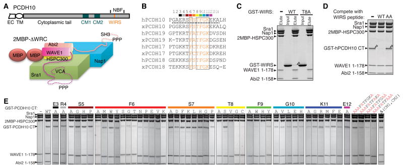
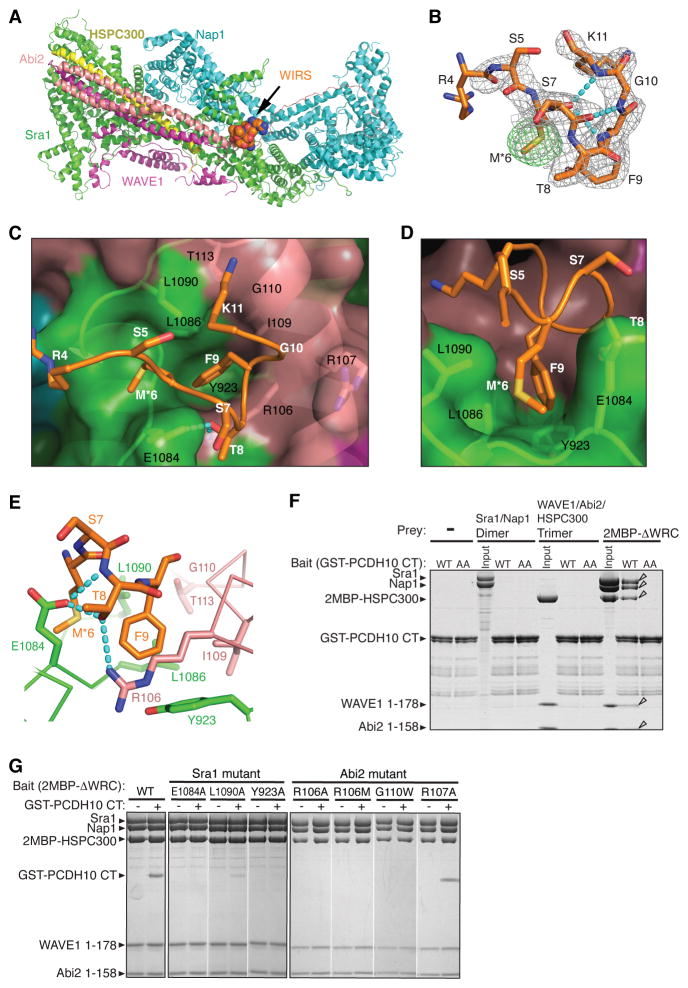
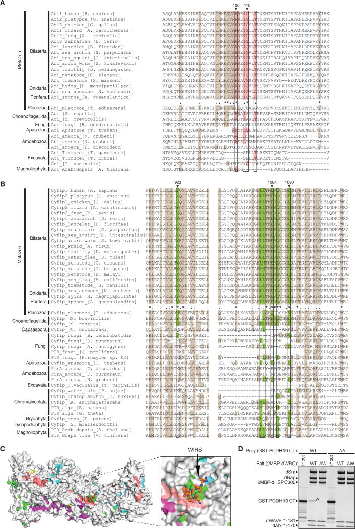
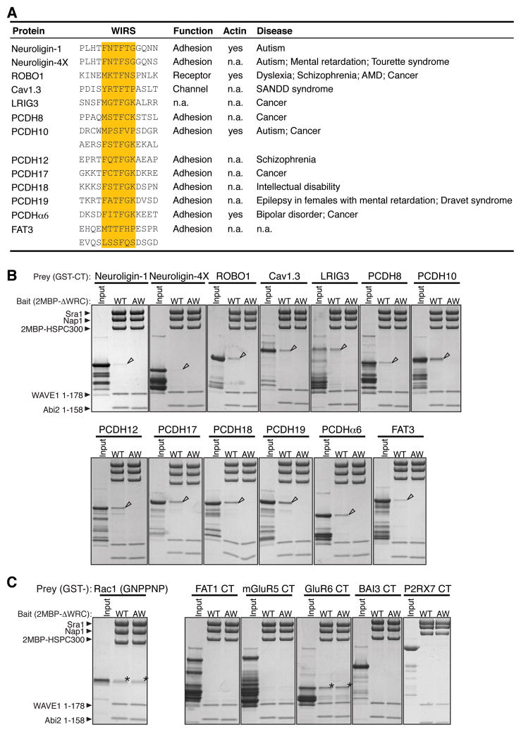
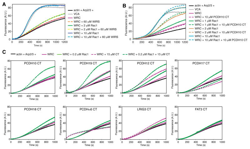
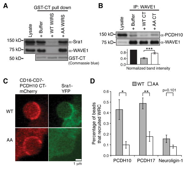
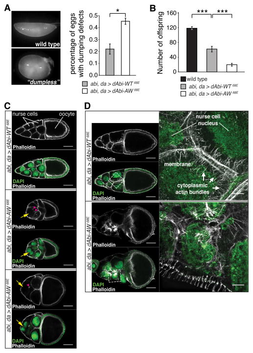
The WAVE regulatory complex (WRC) controls actin cytoskeletal dynamics throughout the cell by stimulating the actin-nucleating activity of the Arp2/3 complex at distinct membrane sites. However, the factors that recruit the WRC to specific locations remain poorly understood. Here, we have identified a large family of potential WRC ligands, consisting of ∼120 diverse membrane proteins, including protocadherins, ROBOs, netrin receptors, neuroligins, GPCRs, and channels. Structural, biochemical, and cellular studies reveal that a sequence motif that defines these ligands binds to a highly conserved interaction surface of the WRC formed by the Sra and Abi subunits. Mutating this binding surface in flies resulted in defects in actin cytoskeletal organization and egg morphology during oogenesis, leading to female sterility. Our findings directly link diverse membrane proteins to the WRC and actin cytoskeleton and have broad physiological and pathological ramifications in metazoans.


Figures

Figure 1
Figure 1. PCDH10 CT Binds to The WRC Using WIRS
(A) Schematic representation of PCDH10 (extracellular domain cropped) and the WRC. (B) Sequence alignment of PCDH10-homologous protocadherin tails (h: human, x: Xenopus tropicalis; dashed line indicates peptide used in crystallography). WIRS is orange, with conserved residues in black boxes. Residues mutated in (E) are color-coded. (C–E) Commassie blue stained SDS-PAGE gels show that immobilized 2MBP-ΔWRC selectively retained GST-WIRS peptide (C), GST-hPCDH10 CT (879–1014) (D), or GST-mPCDH10 CT (778–1014) (E). Triangles indicate bound proteins. In (D), the WIRS peptide, but not a mutant (AA for T8A/F9A), blocked this interaction. In (E), amino acid substitutions are shown below the color-coded wild type residues, or in red letters. See also Figure S1.
Figure 2
Figure 2. WIRS Binds to a Composite Surface Formed by Sra1 and Abi2
(A) Structure of the WIRS/WRC complex (Sra1: green; Nap1: cyan; HSPC300: yellow; Abi2: pink; WAVE1: magenta; WIRS peptide: spheres). (B) 2Fo-Fc electron density map (grey mesh, 1.2 σ) and anomalous scattering map (green mesh, 4 σ) around the WIRS peptide. Cyan dotted lines show intra-peptide hydrogen bonds. (C and D) Top and side views of a semi-transparent WRC surface (key side chains shown under the surface, black labels) with WIRS peptide (white labels). (E) WRC-WIRS interactions; dotted lines show intermolecular hydrogen bonds. (F and G) Commassie blue stained SDS-PAGE gels of eluted proteins are shown. In (F), immobilized GST-PCDH10 CT wild type (WT) or mutant (AA for T1002A/F1003A) selectively retained WRC but not indicated sub-complexes. Open triangles indicate bound proteins. In (G), immobilized 2MBP-ΔWRC mutants selectively retained GST-PCDH10 CT. See also Figure S2 and Table S1.
Figure 3
Figure 3. WIRS Binding Surface is Highly Conserved
(A–B) Sequence alignments of representative organisms. Surface residues of the WIRS binding site (black boxes) are highlighted with pink for Abi (A) and green for Sra1 (B). Other conserved residues were highlighted with brown. Degrees of conservation up to Porifera are represented with ClustalW symbols (* for no change, : for conserved, . for less conserved changes). Residues whose mutation disrupts WIRS binding in Figure 2 are labeled with black triangles on top. Grey amino acids indicate where sequence insertions in alignment were not shown. (C) Surface conservation of the WRC, with the most conserved surface residues (ConSurf score 9) (Ashkenazy et al., 2010) colored as in Figure 2A and less conserved residues (ConSurf score < 9) in grey. (D) Commassie blue stained SDS-PAGE gel shows that immobilized wild type Drosophila WRC (WT), but not a mutant with a disrupted WIRS binding surface (AW for R118A/G122W-Abi) selectively retained GST-PCDH10 CT (WT), but not a mutant (AA for T1002A/F1003A).
Figure 4
Figure 4. Many WIRS Proteins Bind the WRC
(A) WIRS ligands that bind WRC in pull-down assays, with WIRS highlighted in orange. See Table S2 for references. (B and C) Coomassie blue stained gels show proteins selectively retained by immobilized 2MBP-ΔWRC (WT for wild type, AW for R106A/G110W-Abi2). (B) shows verified cytoplasmic tails of WIRS proteins. (C) shows false positive WIRS ligands. Arrows denote bound proteins. Asterisks indicate protein bound to the WRC independent on the WIRS interaction. GST-Rac1-GMPPNP is a positive control. See also Table S2.
Figure 5
Figure 5. WIRS-Containing Tails have Various Effects on WRC’s Activity
(A–C) Actin assembly assays of WIRS (A), PCDH10 CT (B), or other WIRS-containing tails (C). Reactions contain 4 μM actin (5% pyrene labeled), 10 nM Arp2/3 complex, 100 nM WRC217 or VCA (A and B), or 50 nM FL-WRC (C), and/or Rac1 where indicated.
Figure 6
Figure 6. WIRS Proteins Bind the WRC in Cells
(A) Western blots show endogenous WRC retained from mouse brain lysate by immobilized GST-PCDH10 CT, competed by buffer or 100 μM WIRS peptides (WT for wild type, AA for T8A/F9A). (B) Co-immunoprecipitation of WAVE1 from mouse brain lysate, competed by buffer or 5 mM WIRS peptides (WT for wild type, AA for T8A/F9A; similar results achieved by 500 μM GST-PCDH10 CT, see also Extended Experimental Procedures). Quantification of immunoprecipitated PCDH10 is shown below the corresponding samples (n = 7, p = 0.00003). (C) Representative confocal images showing antibody-coated beads clustering mCherry tagged chimeric CD16-CD7-PCDH10 CT receptor (red) expressed in NIH 3T3 cells stably expressing Sra1-YPet (green). (D) Quantification of images represented in (C) for tails of PCDH10 (n = 6, p = 0.014), PCDH17 (n = 4, p = 0.0045), and Neuroligin1 (n = 6, p = 0.10). Each repeat (n) used ~40 or 20 total beads for the wild type and mutant tails, respectively. Error bars stand for SEM, and p values were calculated by Student’s t-test (*: p < 0.05, **: p < 0.005, ***: p < 0.0005). See also Figure S3.
Figure 7
Figure 7. WIRS/WRC Interaction Regulates Oogenesis in Flies
(A) Left: representative images of Drosophila wild type eggs (top) and eggs with a “dumpless” phenotype. Right: quantification of dumping defects in eggs from flies rescued by either dAbi-WT (n = 3) or dAbi-AW (n = 3). For each genotype, each repeat (n) used ~ 220 eggs on average dissected from 7–10 female flies. (B) Quantification of female fertility. Histogram depicts the number of offspring counted from two females mated to wild type males. Bars represent offspring per cross (n = 25 crosses). (C) Representative confocal images of stage 10A egg chambers stained with phalloidin and DAPI. Genotypes as indicated (scale bars: 50 μm). Yellow arrow: lost cortical actin in nurse cells. Magenta arrowhead: ring canals detached from membranes. (D) Left: representative confocal images of stage 10B egg chambers stained with phalloidin and DAPI. Genotypes as indicated (scale bars: 50 μm). Right: structured illumination microscopy (SIM) images of regions in the white boxes (scale bars: 10 μm). Error bars represent SEM, p values were calculated by the Student’s t-test (*: p < 0.05, ***: p < 0.0005). See also Figure S4.

Comment in

  • The shape of things to come.
    Akin O, Zipursky SL. Akin O, et al. Cell. 2014 Jan 16;156(1-2):13-4. doi: 10.1016/j.cell.2013.12.037. Cell. 2014. PMID: 24439365

References

    1. Adams PD, Afonine PV, Bunkoczi G, Chen VB, Davis IW, Echols N, Headd JJ, Hung LW, Kapral GJ, Grosse-Kunstleve RW, et al. PHENIX: a comprehensive Python-based system for macromolecular structure solution. Acta Crystallogr D Biol Crystallogr. 2010;66:213–221. - PMC - PubMed
    1. Alegado RA, Brown LW, Cao S, Dermenjian RK, Zuzow R, Fairclough SR, Clardy J, King N. A bacterial sulfonolipid triggers multicellular development in the closest living relatives of animals. elife. 2012;1:e00013. - PMC - PubMed
    1. Ashkenazy H, Erez E, Martz E, Pupko T, Ben-Tal N. ConSurf 2010: calculating evolutionary conservation in sequence and structure of proteins and nucleic acids. Nucleic Acids Res. 2010;38:W529–533. - PMC - PubMed
    1. Bastock R, St Johnston D. Drosophila oogenesis. Curr Biol. 2008;18:R1082–1087. - PubMed
    1. Bernadskaya YY, Wallace A, Nguyen J, Mohler WA, Soto MC. UNC-40/DCC, SAX-3/Robo, and VAB-1/Eph Polarize F-Actin during Embryonic Morphogenesis by Regulating the WAVE/SCAR Actin Nucleation Complex. PLoS Genet. 2012;8:e1002863. - PMC - PubMed

Publication types

MeSH terms

Substances