. 2014 Jan 16;156(1-2):208-20.
doi: 10.1016/j.cell.2013.12.009.

Local F-actin network links synapse formation and axon branching

Affiliations

Local F-actin network links synapse formation and axon branching

Poh Hui Chia et al. Cell. .

Abstract

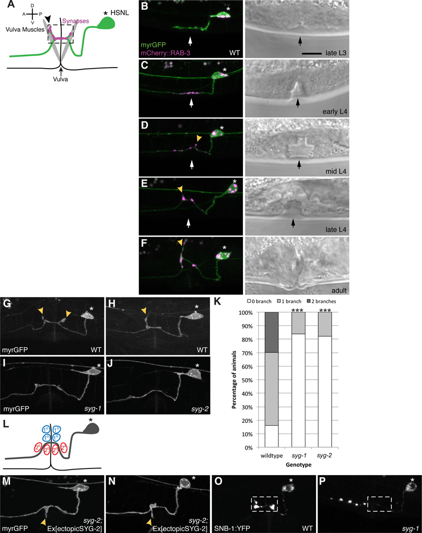
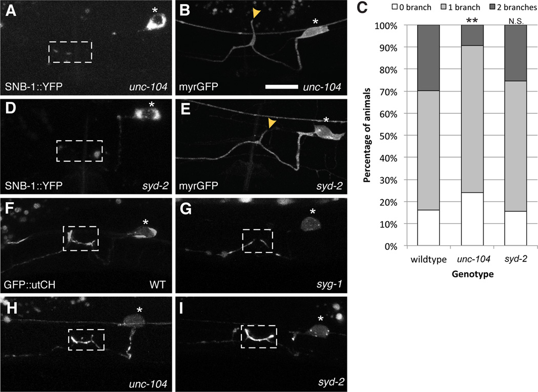
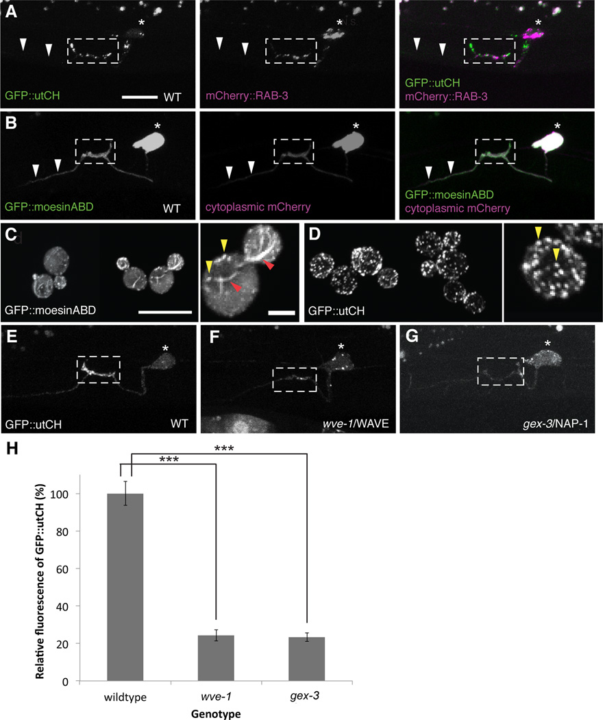
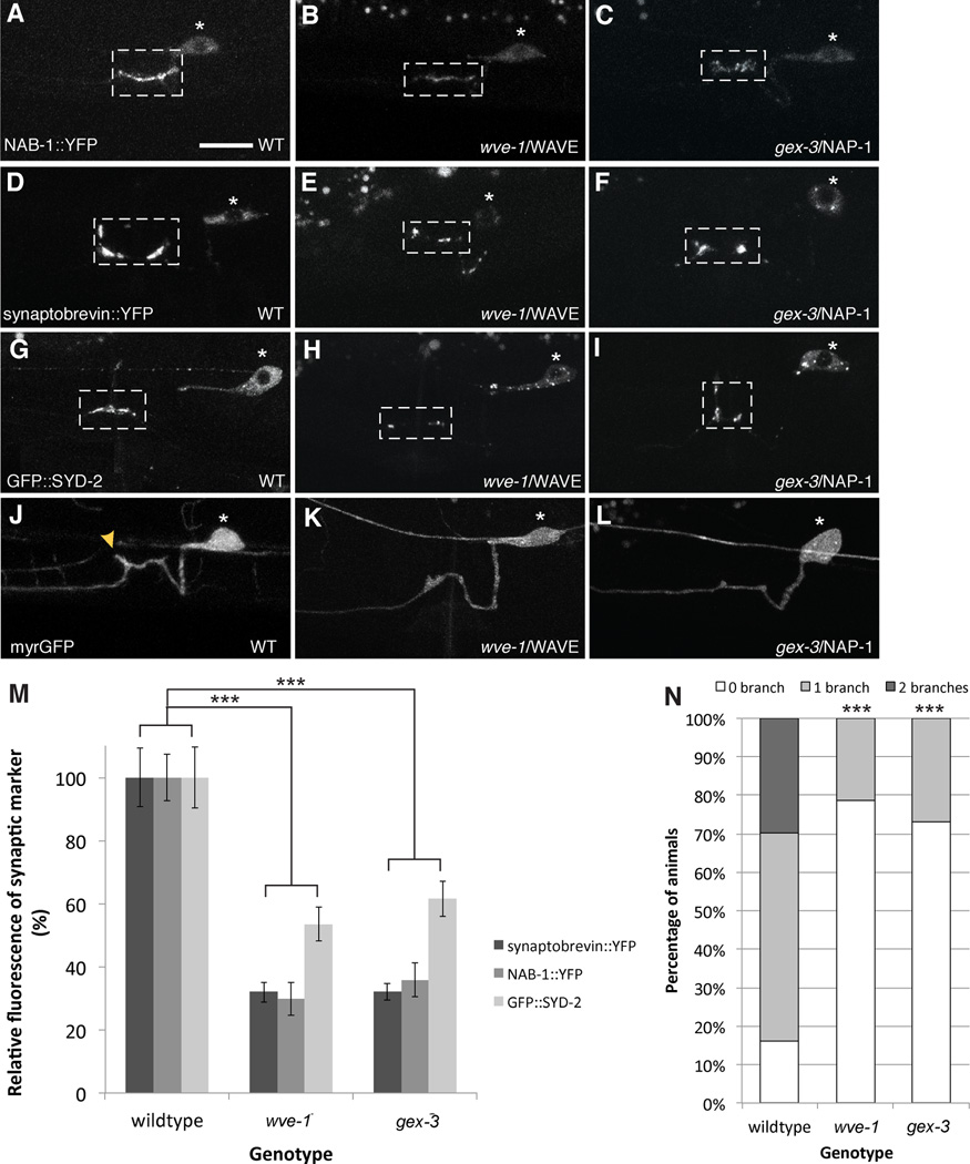
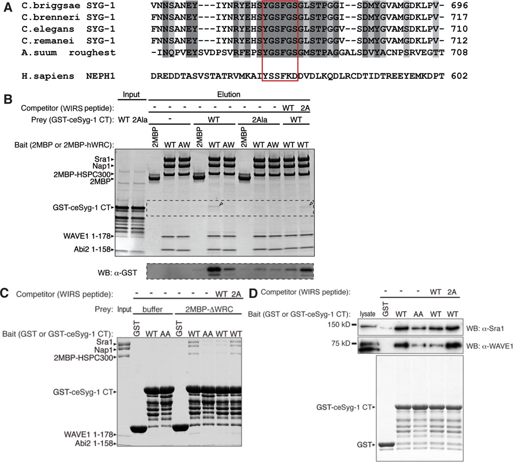
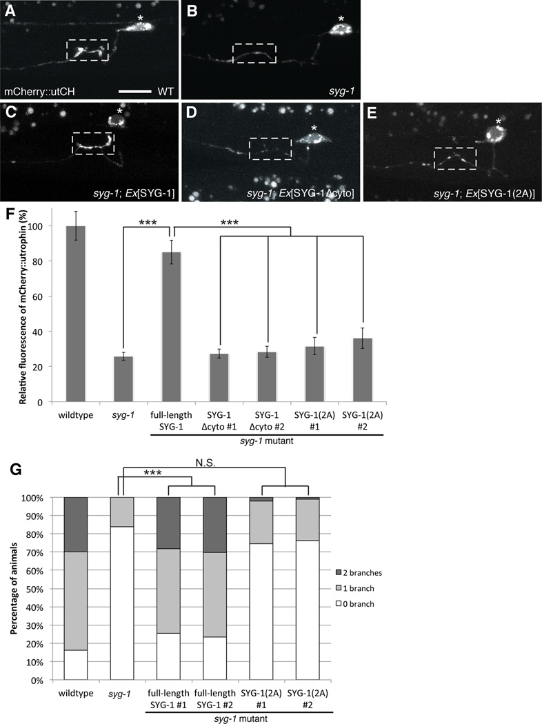
Axonal branching and synapse formation are tightly linked developmental events during the establishment of synaptic circuits. Newly formed synapses promote branch initiation and stability. However, little is known about molecular mechanisms that link these two processes. Here, we show that local assembly of an F-actin cytoskeleton at nascent presynaptic sites initiates both synapse formation and axon branching. We further find that assembly of the F-actin network requires a direct interaction between the synaptic cell adhesion molecule SYG-1 and a key regulator of actin cytoskeleton, the WVE-1/WAVE regulatory complex (WRC). SYG-1 cytoplasmic tail binds to the WRC using a consensus WRC interacting receptor sequence (WIRS). WRC mutants or mutating the SYG-1 WIRS motif leads to loss of local F-actin, synaptic material, and axonal branches. Together, these data suggest that synaptic adhesion molecules, which serve as a necessary component for both synaptogenesis and axonal branch formation, directly regulate subcellular actin cytoskeletal organization.


Figures

Figure 1
Figure 1. Interaction between SYG-1/SYG-2 is required for presynaptic assembly and branch formation
(A) Schematic of HSN. * Denotes the cell body and synapses (pink) form in the synaptic region (dashed box) onto the vulva muscles. Black arrowhead points to axonal collateral branch. (B–F) Representative images depicting the development of HSN neuron. Myristolated GFP highlights the morphology of HSN. Yellow arrowheads point to axonal branches with synapses labeled by mCherry::RAB-3 (pink). Black and white arrows denote the vulva. During the late L3 stage, the main axon is growing across the developing vulva with no visible accumulation of synaptic material (pink). At the early L4 stage, synaptic vesicles begin accumulating at the synaptic region around the vulva. In the mid L4 stage, in some animals, one or two collateral axonal branches extend and become quite pronounced by the late L4 stage. These branches continue to lengthen into the adult stage and accumulate synaptic material, (G, H) Myristolated GFP labels the morphology of HSN. HSN elaborates one or two axonal branches (yellow arrows) that always develop from the synaptic region. (I, J) Branches fail to form in syg-1 or syg-2 mutants. (K) Graph quantifies the percentage of animals in each genotype that elaborates zero, one or two branches. Statistics for each mutant was from comparison with the wildtype values (***p<0.001 with n>100, Fisher’s exact test). (L) Schematic showing the location of primary (1°, red) and secondary (2°, blue) vulva epithelial cells. In wildtype, the 1° vulva epithelial cells express SYG-2. (M, N) Ectopic expression of SYG-2 in 2° vulva cells in syg-2 mutants causes ectopic branches that elaborate ventrally. (O) A wildtype HSN neuron with synapses labeled by synaptobrevin::YFP. (P) syg-1 mutants show ectopic accumulations of synaptobrevin::YFP along the axon anterior to the normal synaptic region around the vulva. Scale bars represent 10 µm.
Figure 2
Figure 2. Synaptic vesicles and active zone proteins are not required for collateral branch formation
(A) In kinesin motor unc-104 mutants, synaptic vesicles labeled by synaptobrevin::YFP fail to get transported to the synaptic region. (B) Loss of unc-104 results in a partial reduction in branch formation. (C) Graph quantifies the percentage of animals in each genotype that elaborate zero, one or two branches. Statistics for each mutant was from comparison with the wildtype values (**p<0.01 with n>100, Fisher’s exact test). (D) syd-2 mutants fail to accumulate synaptic vesicles and active zone molecules (E) but branches are unaffected in syd-2 mutants. (F) GFP::utrophinCH labels synaptic F-actin that is enriched at presynaptic specializations in the L4 stage. (G) This F-actin localization is loss in syg-1 mutants (H, I) but is unaffected in unc-104 and syd-2 mutants. Yellow arrowheads point to collateral branches. Scale bars represent 10 µm. See also Figure S1.
Figure 3
Figure 3. WRC is required for assembling an Arp2/3 mediated actin network at synapses
(A) GFP::utrophinCH labels the F-actin network that is enriched at synapses labeled by mCherry::RAB-3. White arrows point to the anterior axon that has very little GFP::utCH staining. (B) GFP::moesinABD labels the entire HSN neuron (white arrows show bright labeling along the entire axon) with no significant enrichment at presynaptic sites as compared to cytoplasmic mCherry. Scale bars represent 10 µm. (C, D) This difference in actin binding is observed in yeast where GFP::moesinABD binds F-actin cables (red arrowheads) and endocytic F-actin patches (yellow arrowheads) whilst GFP::utCH binds only endocytic F-actin patches Scale bars represent 10 µm and higher magnification image is 2 µm. (E–G) Localization of GFP::utCH at the synaptic region is loss in wve-1 and gex-3 mutants compared to WT. (H) Graph quantifies the average fluorescence intensity for GFP::utCH. wve-1 and gex-3 showed a 66 ± 3% and 67 ± 2% reduction in utCH fluorescence respectively. Each bar represents the average fluorescence value and error bars are ± S.E.M. (***p<0.001 with n>20, Two-tailed Student’s t-test). See also Figure S2 and S3.
Figure 4
Figure 4. WRC is required for both presynapse assembly and axonal branch formation
(A) F-actin dependent active zone protein NAB-1::YFP localizes to synapses in WT animals. Loss of (B) wve-1 or (C) gex-3 results in failed recruitment of NAB-1 to presynaptic sites. (D) A wildtype neuron with synapses labeled by synaptobrevin::YFP. (E, F) wve-1 and gex-3 mutants show partial loss of synaptobrevin::YFP. (G–I) Similarly for active zone molecule SYD-2, wve-1 and gex-3 mutants display a partial reduction in the recruitment of GFP::SYD-2 to synapses. (J) Myristolated GFP highlights the morphology of HSN. (K, L) Most wve-1 and gex-3 mutants fail to extend collateral axonal branches. Yellow arrowheads point to branches. Scale bars represent 10 µm. (M) Graph quantifies the relative average fluorescence of synaptobrevin::YFP, NAB-1::YFP and GFP::SYD-2 in wildtype, wve-1 and gex-3 mutants. Each bar represents the average fluorescence value and error bars are ± S.E.M. (***p<0.001 with n>20, Two-tailed Student’s t-test). See also Figure S4. (N) Graph quantifies the percentage of animals that elaborate zero, one or two branches. wve-1 and gex-3 mutants have significantly less branches as compared to wildtype. Statistics for each mutant was compared against wildtype (***p<0.001 with n>100, Fisher’s exact test). See also Figure S4 and S5.
Figure 5
Figure 5. SYG-1 cytoplasmic tail contains a WRC interacting receptor sequence (WIRS) that specifically bind the WRC
(A) Amino acid sequence alignment of SYG-1/Roughest/NEPH1 homologs. Dark shaded sequences are identical and light shaded sequences are conserved. The WIRS sequence is highlighted in the red box. (B) Pull-down using immobilized di-MBP-tagged human WRC complex as bait (wild type 2MBP-hWRC, or containing R106A/G110W mutations in the Abi2 subunit (AW), which impair binding to WIPS motifs). The top gel is an SDS-PAGE gel stained by coomassie blue and the bottom gel was blotted using mouse anti-GST conjugated to HRP. GST-tagged C. elegans SYG-1 cytoplasmic tail (GST-ceSYG-1 CT) is pulled down by MBP-hWRC (band in the coomasie blue gel is highlighted by black arrow). Making the AW mutation in hWRC interface that interferes with WIRS binding decreases this binding. Mutating the WIRS sequence in GST-ceSYG-1 CT (2Ala) also decreases the binding efficiency. Competitors were chemically synthesized 15 amino acid WIRS peptides (WT for wildtype and 2A for the mutant peptide) and only the wildtype peptide was able to compete for binding. (C) Coommasie blue stained gel from pull-down using immobilized GST or GST-ce-SYG-1 CT as bait and 2MBP-hWRC as prey (wild type and mutants as in panel B), with or without WIRS peptide competitor. The hWRC complex is pulled down by GST-ceSYG-1 CT (D) Pull-down using immobilized GST or GST-ce-SYG-1 CT as bait (wild type and mutants as in panels B and C) and mouse brain lysate as prey, with or without WIRS peptide competitor. Top two gels are western blots with anti-rabbit Sra1 and anti-mouse WAVE1 antibodies respectively; bottom gel is coomassie blue stained to show preys. See also Figure S6.
Figure 6
Figure 6. Local F-actin assembly requires interaction between SYG-1 WIRS and the WRC
Structure-function analysis of SYG-1 (A) mCherry::utCH labels synaptic F-actin in wildtype worms. (B) This enrichment is lost in syg-1 mutants. (C) This defect is rescued by HSN-specific expression of a transgene carrying full-length SYG-1. (D) Expression of SYG-1 lacking its cytoplasmic tail SYG-1Δcyto, fails to rescue. (E) Similarly, expression of SYG-1 with two alanine mutations SYG-1(2A) in the WIRS sequence fails to restore F-actin localization to synapses. Scale bars represent 10 µm. (F) Graph quantifies the relative average fluorescence intensity of mCherry::utCH. Each bar represents the average fluorescence value and error bars are ± S.E.M. For lines expressing SYG-1Δcyto and SYG-1(2A) transgenes, two independent lines were quantified. (***p<0.001 with n>25, Two-tailed Student’s t-test). (G) Graph quantifies the percentage of animals that elaborate zero, one or two branches. Statistics for each mutant was compared against the wildtype values. (***p<0.001 with n>100, Fisher’s exact test). See also Figure S6

Comment in

  • The shape of things to come.
    Akin O, Zipursky SL. Akin O, et al. Cell. 2014 Jan 16;156(1-2):13-4. doi: 10.1016/j.cell.2013.12.037. Cell. 2014. PMID: 24439365

Similar articles

Cited by

References

    1. Besse F, Mertel S, Kittel RJ, Wichmann C, Rasse TM, Sigrist SJ, Ephrussi A. The Ig cell adhesion molecule Basigin controls compartmentalization and vesicle release at Drosophila melanogaster synapses. The Journal of Cell Biology. 2007;177:843–855. - PMC - PubMed
    1. Bleckert A, Photowala H, Alford S. Dual pools of actin at presynaptic terminals. J Neurophysiol. 2012;107:3479–3492. - PMC - PubMed
    1. Bloom O, Evergren E, Tomilin N, Kjaerulff O, Löw P, Brodin L, Pieribone VA, Greengard P, Shupliakov O. Colocalization of synapsin and actin during synaptic vesicle recycling. The Journal of Cell Biology. 2003;161:737–747. - PMC - PubMed
    1. Chen SX, Tari PK, She K, Haas K. Neurexin-Neuroligin Cell Adhesion Complexes Contribute to Synaptotropic Dendritogenesis via Growth Stabilization Mechanisms In&nbsp;Vivo. Neuron. 2010a;67:967–983. - PubMed
    1. Chen Z, Borek D, Padrick SB, Gomez TS, Metlagel Z, Ismail AM, Umetani J, Billadeau DD, Otwinowski Z, Rosen MK. Structure and control of the actin regulatory WAVE complex. Nature. 2010b;468:533–538. - PMC - PubMed

Publication types