. 2014 Jan 16;156(1-2):221-35.
doi: 10.1016/j.cell.2013.11.045.

Tachykinin-expressing neurons control male-specific aggressive arousal in Drosophila

Affiliations

Tachykinin-expressing neurons control male-specific aggressive arousal in Drosophila

Kenta Asahina et al. Cell. .

Abstract

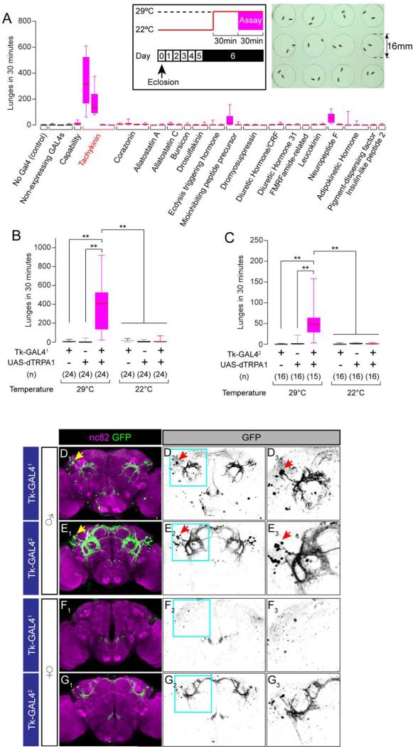
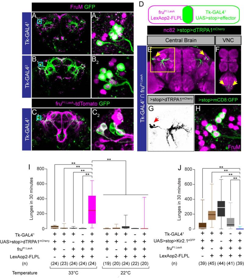
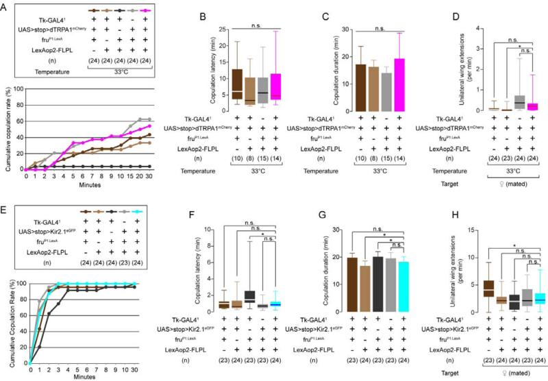
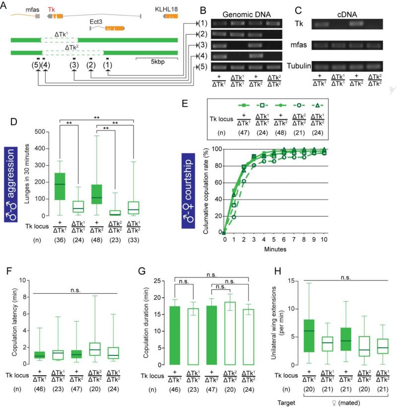
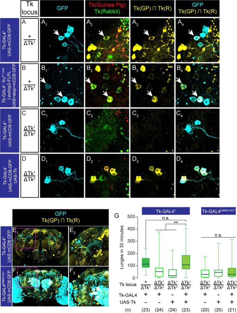
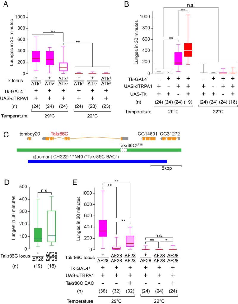
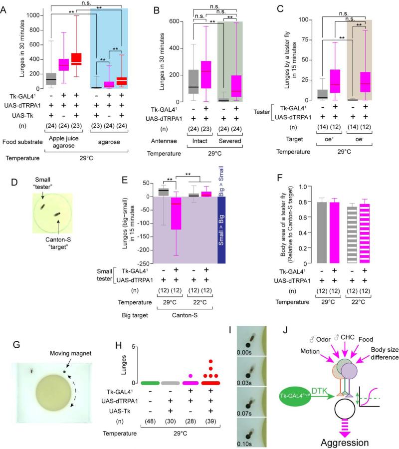
Males of most species are more aggressive than females, but the neural mechanisms underlying this dimorphism are not clear. Here, we identify a neuron and a gene that control the higher level of aggression characteristic of Drosophila melanogaster males. Males, but not females, contain a small cluster of FruM(+) neurons that express the neuropeptide tachykinin (Tk). Activation and silencing of these neurons increased and decreased, respectively, intermale aggression without affecting male-female courtship behavior. Mutations in both Tk and a candidate receptor, Takr86C, suppressed the effect of neuronal activation, whereas overexpression of Tk potentiated it. Tk neuron activation overcame reduced aggressiveness caused by eliminating a variety of sensory or contextual cues, suggesting that it promotes aggressive arousal or motivation. Tachykinin/Substance P has been implicated in aggression in mammals, including humans. Thus, the higher aggressiveness of Drosophila males reflects the sexually dimorphic expression of a neuropeptide that controls agonistic behaviors across phylogeny.


Figures

Figure 1
Figure 1. Tk-GAL4 Lines Label Aggression-promoting Neurons
(A) Number of lunges (boxplot) during dTRPA1-mediated thermogenetic activation of neurons labeled by 42 neuropeptide-GAL4 drivers. A box indicates lower quartile, median and higher quartile, from bottom to top. Whiskers represent the range of the remaining data points. Names of the genes from which the promoter fragments were generated are listed below plots. n = 10-24. The inlet illustrates the experimental design (left) and the 12-well aggression chamber used in this assay (right). (B, C) Number of lunges (boxplot) during thermogenetic activation of Tk-GAL41(B) and Tk-GAL42(C) neurons. Genotypes, number of pairs and temperature tested are indicated below the plot. ** p < 0.01 (Kruskal-Wallis and post-hoc Mann-Whitney U-tests). Horizontal bar above 22°C data indicates pooling for statistical analysis (Kruskal-Wallis test, p > 0.05). (D-G) Brains of Tk-GAL41; UAS-mCD8:GFP male (D), female (F), Tk-GAL42; UAS-mCD8:GFP male (E) and female (G) immunostained with anti-GFP antibody (green) and the neuropil marker nc82 (magenta) (D1-G1). D2-G2: GFP only. D3-G3: region within a cyan square in D2-G2 (magnified). Arrows: the lateral cluster neurons (see text). Also see Supplemental Figure S1 and Supplemental Movie S1.
Figure 2
Figure 2. Tk-GAL4FruM neurons control male-male aggression
(A, B) Tk-GAL41; UAS-mCD8:GFP (A) and Tk-GAL42; UAS-mCD8:GFP (B) male brains immunostained with anti-GFP antibody (green) and anti-FruM antisra (magenta). A2, B2: region within a cyan square in A1 and B1 (magnified). (C) Tk-GAL41; fruP1.LexA/ UAS-mCD8:GFP, LexAop2-tdTomato male brain immunostained with anti-GFP antibody (green) and anti-DsRed antibody (magenta). C2: region within a cyan square in C1 (magnified). (D) The schematic of the genetic intersectional strategy utilized in this study. (E, F) Tk-GAL4FruM neurons in the brain (E) and VNC (F) immunostained with anti-DsRed antibody (green) and nc82 (magenta). Arrows: cell bodies stained with anti-DsRed antibody. (G) Region within a cyan square in (E) (magnified). Only the image of anti-DsRed is shown. Arrow: cell bodies stained with anti-DsRed antibody. (H) Four Tk-GAL4FruM neurons in a male brain, immunostained with anti-GFP antibody (green) and anti-FruM antisera (magenta). The magnification compares to images A2, B2 and C2. (I, J) Number of lunges during thermogenetic activation (I) and silencing (J) of Tk-GAL4FruM neurons. For (I) and (J), ** p < 0.01 (Kruskal-Wallis and post-hoc Mann-Whitney U-tests). Also see Supplemental Figure S2 and Supplemental Movie S2.
Figure 3
Figure 3. Tk-GAL4FruM neurons do not modulate courtship behavior
(A) Cumulative copulation curve for males during thermogenetic activation of Tk-GAL4FruM neurons. Tk-GAL41; fruP1.LexA/ LexAop2-FLPL control males were eliminated from subsequent analyses. (B-D) Copulation latency (B), duration (C, mean ± S.D.) and unilateral wing extension frequency (D) during thermogenetic activation of Tk-GAL4FruM neurons. (E) Cumulative copulation curve for males during silencing of Tk-GAL4FruM neurons. (F-H) Copulation latency (F), duration (G) and unilateral wing extension frequency (H) during silencing of Tk-GAL4FruM neurons. For (B), (D), (F) and (H), * p < 0.05, n.s.: p > 0.05 (Kruskal-Wallis and post-hoc Mann-Whitney U-tests. For (C) and (G), * p < 0.05, n.s.: p > 0.05 (one-way ANOVA or one-way ANOVA and post-hoc Student's t-tests). Also see Supplemental Figure S3.
Figure 4
Figure 4. Null mutations on Tk specifically affect male-male aggression
(A) Schematic view of the Tk gene locus and deletions by ΔTk1 and ΔTk2. Black bars (1)-(5) represent the regions targeted in the PCR analysis in (B). (B) PCR analysis against regions (1)-(5) in (A) from genomic DNA samples of Tk deletion mutants. (C) RT-PCR targeted to the Tk, mfas and Tubulin (α-Tubulin at 84B) (positive control) gene transcripts from cDNA samples of Tk deletion mutants. (D-H) Number of lunges (D), cumulative copulation curve (E), copulation latency (F), copulation duration (G) and unilateral wing extension frequency (H) performed by Tk deletion mutants. For (D), (F) and (H), ** p < 0.01, n.s.: p > 0.05 (Kruskal-Wallis or Kruskal-Wallis and post-hoc Mann-Whitney U-tests). For (G), n.s.: p > 0.05 (one-way ANOVA and post-hoc Student's t-test). Also see Supplemental Figure S4.
Figure 5
Figure 5. Tk gene products in the Tk-GAL4FruM neurons are sufficient to maintain normal level of aggression
(A-D) Tk-GAL41 lateral cluster neurons in +/ΔTk1 (A), ΔTk2/ΔTk1 (C), ΔTk2/ΔTk1 plus UAS-Tk (D) backgrounds, and Tk-GAL4FruM neurons in the +/ΔTk1 (B) background, immunostained with anti-GFP antibody (cyan; A1-D1, A4-D4), anti-DTK guinea pig antiserum (red, A2-D2) and anti-DTK rabbit antiserum (green, A2-D2). The overlap of the two antisera is shown in yellow in A3-D3 and A4-D4. Arrows: the GFP+, DTK+ neurons. (E,F) Tk-GAL41 (E) and Tk-GAL4GMR61H07 (F) neurons in male brains immunostained with anti-GFP antibody (cyan), anti-Tk guinea pig antiserum and anti-Tk rabbit antiserum (shown as the overlap in yellow). (E2, F2): region within a magenta square in E1 and F1 (magnified). (G) Number of lunges in ΔTk1/ΔTk2 rescued by Tk-GAL41 or Tk-GAL4GMR61H07 driving UAS-Tk. Left: ** p < 0.01, n.s. p > 0.05 (Kruskal-Wallis and post-hoc Mann-Whitney U-tests) Right: n.s.: Kruskal-Wallis test, p > 0.05. Also see Supplemental Figure S5.
Figure 6
Figure 6. The Tk system gates the aggression-promoting effect of Tk-GAL41 neurons
(A, B) Number of lunges during thermogenetic activation of Tk-GAL41 neurons in the Tk null mutant background (A), or with Tk overexpression (B). (C) Schematic of the Takr86C gene locus, its deletion in Takr86C F28 and the region covered by p[acman] CH322-17N40. (D) Number of lunges performed by Takr86CΔF28 mutants. (E) Number of lunges during thermogenetic activation of Tk-GAL41 neurons in the Takr86CΔF28 mutant background. For (A), (B), (D) and (E), ** p < 0.01, * p < 0.05, n.s.: p > 0.05 (Kruskal-Wallis and/or post-hoc Mann-Whitney U-tests). Also see Supplemental Figure S6.
Figure 7
Figure 7. Activation of Tk-GAL41 neurons overrides the absence of aggression-promoting cues
(A-C) Number of lunges during thermogenetic activation of Tk-GAL41 neurons, (A) on an apple juice agarose (left) or pure agarose (right) substrate; (B) with intact (left) or surgically removed (right) antennae; and (C) toward oe+ (left) or oe− (right) “target” males. (D) Example image of a normal-sized “target” Canton-S male and a small “tester” male fly (70% the size of the target fly). (E) Lunge number difference between smaller tester and larger target flies. Values in the shaded area indicate that the smaller testers performed more lunges than the larger targets. (F) Relative body sizes (mean ± S.D.) of “tester”vs. target flies. (G) Image of the “moving magnet” setup. (H) Number of lunges (scatter plot) toward the magnet during thermogenetic activation of Tk-GAL41 neurons. Total recording time per session was 4 minutes (see Experimental Procedures). (I) Example of a lunge performed by a male Tk-GAL41, UAS-dTRPA1; UAS-Tk fly toward a magnet. (J) Schematic illustrating possible influence of Tk-GAL4FruM neurons in the male fly brain in relation to the processing of sensory cues regulating aggression. For (A-C) and (E), ** p < 0.01, * p < 0.05 (Kruskal-Wallis and post-hoc Mann-Whitney U-tests). For (F), n.s.: p > 0.05 (one-way ANOVA and post hoc Student's t-test. Also see Supplemental Movie S3.

Comment in

References

    1. Alekseyenko OV, Chan YB, Li R, Kravitz EA. Single dopaminergic neurons that modulate aggression in Drosophila. Proc Natl Acad Sci U S A. 2013;110:6151–6156. - PMC - PubMed
    1. Alekseyenko OV, Lee C, Kravitz EA. Targeted manipulation of serotonergic neurotransmission affects the escalation of aggression in adult male Drosophila melanogaster. PLoS One. 2010;5:e10806. - PMC - PubMed
    1. Baier A, Wittek B, Brembs B. Drosophila as a new model organism for the neurobiology of aggression? J Exp Biol. 2002;205:1233–1240. - PubMed
    1. Bargmann CI. Beyond the connectome: how neuromodulators shape neural circuits. Bioessays. 2012;34:458–465. - PubMed
    1. Bentley D, Konishi M. Neural control of behavior. Annu Rev Neurosci. 1978;1:35–59. - PubMed

Publication types

LinkOut - more resources