. 2014 Jan 15;15(1):95-102.
doi: 10.1016/j.chom.2013.12.010.

Gut dysbiosis promotes M2 macrophage polarization and allergic airway inflammation via fungi-induced PGE₂

Affiliations

Gut dysbiosis promotes M2 macrophage polarization and allergic airway inflammation via fungi-induced PGE₂

Yun-Gi Kim et al. Cell Host Microbe. .

Abstract

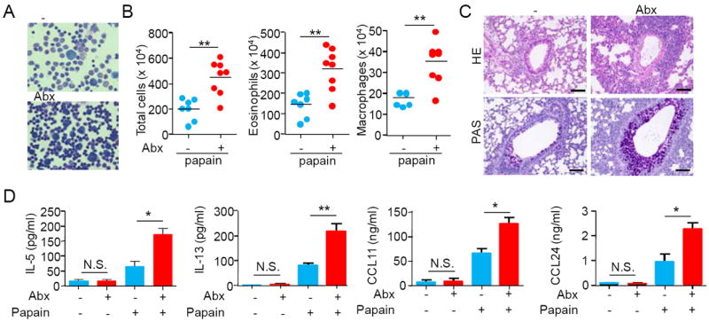
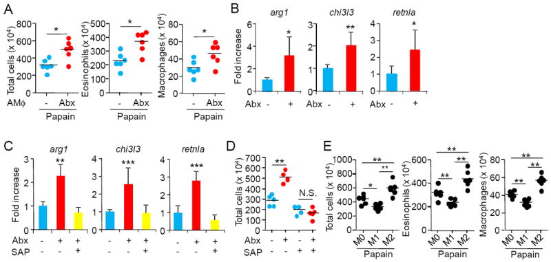
Although imbalances in gut microbiota composition, or "dysbiosis," are associated with many diseases, the effects of gut dysbiosis on host systemic physiology are less well characterized. We report that gut dysbiosis induced by antibiotic (Abx) treatment promotes allergic airway inflammation by shifting macrophage polarization in the lung toward the alternatively activated M2 phenotype. Adoptive transfer of alveolar macrophages derived from Abx-treated mice was sufficient to increase allergic airway inflammation. Abx treatment resulted in the overgrowth of a commensal fungal Candida species in the gut and increased plasma concentrations of prostaglandin E₂ (PGE₂), which induced M2 macrophage polarization in the lung. Suppression of PGE₂ synthesis by the cyclooxygenase inhibitors aspirin and celecoxib suppressed M2 macrophage polarization and decreased allergic airway inflammatory cell infiltration in Abx-treated mice. Thus, Abx treatment can cause overgrowth of particular fungal species in the gut and promote M2 macrophage activation at distant sites to influence systemic responses including allergic inflammation.


Figures

Figure 1
Figure 1. Antibiotics treatment exacerbates allergic airway inflammation
(A-D) Antibiotic (clindamycin + cefoperazone) (Abx)-treated or untreated (-) mice were challenged intranasally with papain every 3 days for a total of five times. One day after the final challenge, mice were analyzed for morphology of BAL cells (Wright’s stain, original magnification × 1000) (A), total and differentiated cell counts in BAL (B), lung histology (HE or PAS stain) (scale bar, 100 μm) (C), and cytokine concentration in BAL fluids (D). Representative results in each group of A (n=7 or 8) and C (n=4~6) are shown. Error bars indicate SD (D) (n=7). Results are representative of three independent experiments (B and D). *P < 0.05; **P < 0.01. N.S. denotes not significant. See also Figure S1.
Figure 2
Figure 2. Antibiotic treatment induces M2 macrophage polarization
(A) Mice were adoptively transferred i.n. with alveolar macrophages (AMϕs) purified from untreated (-) or Abx-treated mice, and challenged with papain and analyzed for BAL cells, as described in Fig 1. (B) AMϕs purified from untreated (-) or Abx-treated (+) mice were subjected to real-time RT-PCR (n=5). (C) Abx-treated (+) or untreated (-) mice were administered i.n. with SAP (+) or PBS (-) every 3 days for a total of five times. One day after the final SAP treatment, mice were analyzed for expression of M2 macrophage marker genes in AMϕs (n=5). (D) Mice were left untreated (-) or treated with Abx and were challenged with papain every 3 days for a total of five times. The mice were also injected i.n. with serum amyloid P (SAP) or PBS one day before each papain challenge every 3 days for a total of five times. One day after the final challenge with papain, mice were analyzed for BAL cells, as described in Fig 1. (E) Mice were adoptively transferred i.n. with bone marrow-derived cultured macrophages (M0), M1 macrophages, or M2 macrophages, and then challenged i.n. with papain and analyzded for BAL cells, as described in Fig 1. Results are representative of three independent experiments. * P < 0.05; **P < 0.01, ***P < 0.001. N.S. denotes not significant. Error bars indicate SD. See also Figure S2.
Figure 3
Figure 3. Antibiotic treatment facilitates fungal overgrowth in the gut, which induces M2 macrophage polarization
(A–C) Mice were left untreated or treated orally with Abx (CLM: clindamycin; CPZ: cefoperazone; ABPC: ampicillin; MNZ: metronidazole; STM: streptomycin) for 2 weeks and analyzed for fungal colonization in the feces (A). Mice were then challenged i.n. with papain and analyzed for BAL cells, as described in Fig 1 (B). Correlation between fungal colonization and total cell number in BAL were analyzed (C). (D, E) Cecum sections in mice untreated (-) or treated with Abx (clindamycin + cefoperazone) for 2 weeks were stained with PAS (D) or DAB (E), as described in supplemental materials. Arrows indicate Candida in the lamina propria. Scale bar, 10 μm. (F-H) Mice were left untreated or treated orally with either Abx (clindamycin + cefoperazone) or Abx plus 5-FC for 2 weeks and analyzed for fungal colonization in the feces (F). AMϕs were subjected to real-time RT-PCR (G) (n=5). Mice were then challenged i.n. with papain and analyzed for BAL cell number, as described in Fig 1 (H). Results are representative of three independent experiments. *P < 0.05; **P< 0.01. N.S. denotes not significant. See also Figure S3.
Figure 4
Figure 4. Elevated PGE2 levels associated with intestinal fungal overgrowth contribute to M2 polarization and increase of allergic airway inflammatory cell infiltration
(A) Peripheral blood monocytes purified from untreated (-) or Abx (clindamycin + cefoperazone)-treated mice were subjected to real-time RT-PCR (n=5). (B) Candida parapsilosis (Cp) isolated from mouse feces was cultured or not for 48 h in the presence or absence of 500 μM arachidonic acid (AA) and assayed for prostaglandin E2(PGE2) metabolite (PGEM) in the culture supernatant. (C) Mice were left untreated or treated orally with either Abx, or Abx plus 5-FC for 2 weeks and analyzed for serum PGEM levels. (D) Abx-treated or untreated (-) mice were challenged i.n. with PBS or papain every 3 days for a total of five times. One day after the final challenge, mice were analyzed for PGEM concentration in BAL fluids. (E-G) Ptges+/+ (E-G) or Ptges-/- (E) mice were left untreated or treated orally with either Abx, aspirin (Asp) or both for 2 weeks and analyzed for serum PGEM levels (E), expression of M2 macrophage marker genes in AMϕs by real-time RT-PCR (F) (n=5). The mice were then challenged i.n. with papain and analyzed, as described in Fig 1 (G). (H) Mice were left untreated or treated orally with either Abx, celecoxib or both for 2 weeks and then challenged i.n. with papain and analyzed, as described in Fig 1. (I) Mice were intraperitoneally injected with dimethyl PGE2 (Dm-PGE2) or PBS every day for a total of five times. One day after the final challenge, mice were analyzed for expression of M2 macrophage marker genes in AMϕs (n=5). (J) Mice were challenged i.n. with papain every 3 days for a total of five times. The mice were also injected i.p. with dimethyl PGE2 (Dm-PGE2) or PBS one day before each papain challenge for a total of five times. One day after the final challenge with papain, mice were analyzed for BAL cells, as described in Fig 1. (K) AMϕs purified from naive mice were cultured in the presence or absence of PGE2 for 24 h and transferred i.n. to naïve mice. The mice were then challenged i.n. with papain and analyzed for BAL cells, as described in Fig 1. Results are representative of three independent experiments. *P < 0.05; **P < 0.01. Error bars indicate SD. N.S. denotes not significant. See also Figure S4.

Comment in

References

    1. Abt MC, Osborne LC, Monticelli LA, Doering TA, Alenghat T, Sonnenberg GF, Paley MA, Antenus M, Williams KL, Erikson J, et al. Commensal bacteria calibrate the activation threshold of innate antiviral immunity. Immunity. 2012;37:158–170. - PMC - PubMed
    1. Alem MA, Douglas LJ. Effects of aspirin and other nonsteroidal anti-inflammatory drugs on biofilms and planktonic cells of Candida albicans. Antimicrob Agents Chemother. 2004;48:41–47. - PMC - PubMed
    1. Arumugam M, Raes J, Pelletier E, Le Paslier D, Yamada T, Mende DR, Fernandes GR, Tap J, Bruls T, Batto JM, et al. Enterotypes of the human gut microbiome. Nature. 2011;473:174–180. - PMC - PubMed
    1. Boulet L, Ouellet M, Bateman KP, Ethier D, Percival MD, Riendeau D, Mancini JA, Methot N. Deletion of microsomal prostaglandin E2 (PGE2) synthase-1 reduces inducible and basal PGE2 production and alters the gastric prostanoid profile. J Biol Chem. 2004;279:23229–23237. - PubMed
    1. Buslau M, Menzel I, Holzmann H. Fungal flora of human faeces in psoriasis and atopic dermatitis. Mycoses. 1990;33:90–94. - PubMed

Publication types

MeSH terms