. 2014 Feb 20;40(2):225-34.
doi: 10.1016/j.immuni.2013.12.011. Epub 2014 Jan 16.

Costimulatory molecule DNAM-1 is essential for optimal differentiation of memory natural killer cells during mouse cytomegalovirus infection

Affiliations

Costimulatory molecule DNAM-1 is essential for optimal differentiation of memory natural killer cells during mouse cytomegalovirus infection

Tsukasa Nabekura et al. Immunity. .

Abstract

Recent studies demonstrate that natural killer (NK) cells have adaptive immune features. Here, we investigated the role of the costimulatory molecule DNAM-1 in the differentiation of NK cells in a mouse model of cytomegalovirus (MCMV) infection. Antibody blockade of DNAM-1 suppressed the expansion of MCMV-specific Ly49H(+) cells during viral infection and inhibited the generation of memory NK cells. Similarly, DNAM-1-deficient (Cd226(-/-)) Ly49H(+) NK cells exhibited intrinsic defects in expansion and differentiation into memory cells. Src-family tyrosine kinase Fyn and serine-threonine protein kinase C isoform eta (PKCη) signaling through DNAM-1 played distinct roles in the generation of MCMV-specific effector and memory NK cells. Thus, cooperative signaling through DNAM-1 and Ly49H are required for NK cell-mediated host defense against MCMV infection.


Figures

Figure 1
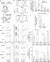
Figure 1. Phenotype of DNAM-1+ and DNAM-1 NK cells
(A) Expression of DNAM-1 on NK cells in the spleen, blood, BM, and liver, gating on TCRβNK1.1+ lymphocytes. Thin and bold lines represent staining with control Ig and anti-DNAM-1 mAb. (B) Developmental stages of DNAM-1+ and DNAM-1 NK cells as determined by co-staining with CD27 and CD11b, gating on TCRβNK1.1+ lymphocytes. (C) Ly49H expression on DNAM-1+ (thick lines) and DNAM-1 (thin lines) NK cells, gated on TCRβNK1.1+ lymphocytes. Percentages of Ly49H+ NK cells in DNAM-1+ (bold numbers) and DNAM-1 NK (thin numbers) cells are shown. (D) Expression of Ly49C and/or Ly49I on DNAM-1+ (thick lines) and DNAM-1 (thin lines) NK cells. Data are representative of 2–4 experiments. (E) IFN-γ production and degranulation of Ly49H+DNAM-1+ and Ly49H+DNAM-1 NK cells after co-culture with RMA cells expressing m157, CD155, or m157 and CD155. Data are representative of 2 independent experiments (n = 3–6 per experiment). (F) Twenty-five million WT splenocytes were transferred into DAP12-deficient mice and infected with 1 × 10⁵ pfu MCMV. IFN-γ production by Ly49H+ NK cells was analyzed by intracellular staining on day 1.5 pi. Percentages of IFN-γ+Ly49H+ NK cells and mean fluorescent intensity (MFI) of IFN-γ of Ly49H+ NK cells within the DNAM-1+ and DNAM-1 Ly49H+ NK cell subsets are shown. Data are representative of 3 experiments (n = 3–4 mice per experiment). Error bars show s.e.m. *p <0.05.
Figure 2
Figure 2. DNAM-1 antibody blockade suppresses the NK cell response to MCMV
(A) WT mice were inoculated with control Ig or a neutralizing mAb against DNAM-1 on the day before infection with 5 × 10⁵ pfu MCMV. Viral burden in the liver and spleen was measured on day 3 pi. (B) One hundred thousand WT Ly49H+ NK cells were transferred into DAP12-deficient mice and infected with 1 × 10⁵ pfu MCMV. Mice were inoculated with 100 µg control Ig or anti-DNAM-1 mAb on the day before infection and day 3 pi. The absolute number of Ly49H+ NK cells per ml blood. (C) DAP12-deficient mice receiving 1 × 10⁵ WT Ly49H+ NK cells were infected with MCMV, and treated with 100 µg control Ig or anti-DNAM-1 mAb on days 7, 14, and 21 pi. The absolute number of Ly49H+ NK cells per ml blood and percentages of Ly49H+ memory NK cells in the spleen on day 28 pi. Data were pooled from 2 experiments (n = 6 mice per mAb group). Error bars show s.e.m. *p <0.05, **p <0.01. See also Figure S1.
Figure 3
Figure 3. DNAM-1 is required for expansion of effector NK cells and differentiation of memory NK cells during MCMV infection
(A) NK cells were purified from WT and Cd226−/− mixed BM chimeric mice. One hundred fifty thousand WT and Cd226−/− Ly49H+ NK cells were transferred into Ly49H-deficient mice and infected with 1 × 10⁵ pfu MCMV. (B) Activation markers CD69 and KLRG1 on Ly49H+ NK cells at day 1.5 and 7 pi, respectively, and IFN-γ production on day 1.5 pi. Thin and bold lines represent Cd226−/− Ly49H+ and WT Ly49H+ NK cells. (C) Phenotype of WT and Cd226−/− naïve, effector (day 7 pi), and memory (day 25 pi) NK cells. Expression of KLRG1, CD11b, CD27, and Ly6C on Ly49H+ NK cells is shown. (D) The kinetics of the absolute number of Ly49H+ NK cells in blood were represented as the ratio relative to the number of Ly49H+ NK cells in the blood on day 0 (before infection). Data are representative of 3 experiments (n = 6–7 mice). (E) Memory NK cells were isolated from recipient mice 30 days after primary infection, transferred into naïve Ly49H-deficient mice, and infected with MCMV. Secondary expansion of Ly49H+memory NK cells in the spleen on day 7 after infection with MCMV was represented as the fold expansion relative to the number of memory NK cells detected in the spleen of mice adoptively transferred with an aliquot of Ly49H+ memory NK cells but not infected with MCMV. Data are pooled from 2 experiments (n = 6 mice). (F and G) WT and Cd226−/− memory NK cells were purified from recipient mice 25–28 days after primary infection, and WT and Cd226−/− naïve NK cells were purified from BM chimeric mice. Ten thousand WT and Cd226−/− naïve and memory Ly49H+ NK cells were transferred separately into Ly49H-deficient or DAP12-deficient mice and infected with MCMV. The copy number of MCMV IE1 gene in blood on day 3 pi (F) and day 7 pi (G) was analyzed by quantitative PCR. Data were pooled from 2 experiments (n = 6–8 mice per group). *p <0.05, **p <0.01, and ***p <0.005. Error bars show s.e.m. See also Figure S2 and S3.
Figure 4
Figure 4. DNAM-1 signaling is required for optimal differentiation of Ly49H+ NK cells during MCMV infection
(A and B) NK cells were purified from BM chimeric mice reconstituted with WT, Cd226−/−, and DNAM-1-rescued Cd226−/− HPCs. Ly49H+ NK cells were isolated from chimeric mice and transferred into Ly49H-deficient mice, and then infected with 1 × 10⁵ pfu MCMV. The number of Cd226−/− and DNAM-1-rescued Ly49H+ effector NK cells on day 7 pi (A) and memory NK cells on day 25 pi (B) in the spleen was represented as the fold expansion relative to the number of WT Ly49H+ NK cells on days 7 and 25 pi in the spleen. Data are pooled from 2 experiments (n = 4 mice per rescued construct). *p <0.05. (C-F) NK cells were purified from BM chimeric mice of WT and Fyn−/− mice, or WT and Prkch−/− mice. WT and Fyn−/− Ly49H+ NK cells (4 × 10⁵ cells), or WT and Prkch−/−Ly49H+ NK cells (1 × 10⁵ cells) were transferred into Ly49H-deficient mice and infected with MCMV. (C and E) The kinetics of the absolute number of Ly49H+ NK cells in blood were represented as the ratio relative to the number of Ly49H+ NK cells in blood on day 0 (before infection). (D and F) Memory NK cells were transferred into naïve Ly49H-deficient mice and infected with MCMV. Secondary expansion of Ly49H+ memory NK cells in the spleen on day 7 after infection with MCMV was represented as the fold expansion relative to the number of memory NK cells detected in the spleen of mice adoptively transferred with an aliquot of Ly49H+ memory NK cells but not infected with MCMV. Data are representative of 2–3 experiments (n = 3–5). Error bars show s.e.m. *p <0.05. See also Figure S4.
Figure 5
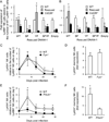
Figure 5. DNAM-1 on Ly49H+ NK cells is modulated during MCMV infection
(A) DNAM-1 on Ly49H+ NK cells in the spleen of mice infected with MCMV on day 7 pi. Data are representative of 2 experiments (n =2–3 in each group). (B) Percentages of DNAM-1-negative NK cells within the Ly49H+ and Ly49H NK cell subsets in the blood of mice infected with 5 × 10⁵ pfu WT or Δm157 MCMV. *p <0.05 vs. the other 3 groups. (C) Twenty-five million CellTrace Violet-labeled splenocytes were transferred into DAP12-deficient mice and infected with 1 × 10⁵ pfu MCMV. Expression of DNAM-1 on Ly49H+ NK cells in the spleen was analyzed on days 3 and 6 pi. Data are representative of 3 experiments (n = 3–4 mice per time point) (B and C). (D and E) One or two hundred thousand CD45.1+Ly49H+ NK cells were transferred into Ly49H-deficient mice and infected with MCMV. (D) MFI of DNAM-1 on Ly49H+ NK cells in the spleen. Data were pooled from 6 experiments (n = 3–5 mice per time point). *p <0.05, **p <0.01 vs. day 0. (E) DNAM-1 on CD45.1+Ly49H+ memory NK cells in the spleen on day 35 pi. Data are representative of more than 5 experiments. (F) Twenty-five million WT splenocytes were transferred into DAP12-deficient mice and infected with MCMV. Cell death of Ly49H+ NK cells was analyzed by Annexin V staining on days 3 and 6 pi. Data are representative of 3 experiments (n = 3–4 mice per experiment). *p <0.05, **p <0.01 vs. DNAM-1-. Error bars show s.e.m. See also Figure S5.
Figure 6
Figure 6. DNAM-1 and DNAM-1+ NK cells expand and differentiate into memory NK cells during MCMV infection
One hundred thousand purified Ly49H+DNAM-1+ or Ly49H+DNAM-1 NK cells were transferred separately into DAP12-deficient mice and infected with 1 × 10⁵ MCMV. Percentages of Ly49H+DNAM-1+ and Ly49H+DNAM-1 NK cells in blood are shown. Data are representative of 3 experiments (n = 4–5 mice). Error bars show s.e.m.
Figure 7
Figure 7. DNAM-1 ligands are upregulated after MCMV infection in vivo
(A) WT mice were infected with 1 × 10⁶ pfu MCMV. Expression of CD155 and CD112 on hematopoietic cells in the spleen of infected mice on days 1 and 3 pi was analyzed by flow cytometry. NK cells (TCRβNK1.1+), NKT cells (TCRβ+NK1.1+), CD4+ T cells (TCRβ+CD4+), CD8+ T cells (TCRβ+CD8α+), B cells (CD11bB220+), DCs (CD11chighI-AE+), granulocytes (Gr; CD11b+Gr-1high), macrophages (Mφ; CD11b+Gr-1low) were assessed. (B) Cre-dependent YFP reporter mice were infected with 2 × 10⁴ pfu Cre-MCMV. YFP+ hematopoietic cells in the spleen of infected mice on days 1 and 3 pi were analyzed. Data are representative of 2 experiments (n = 3 mice per time point) (A and B). *p <0.05 vs. uninfected. (C) WT mice were infected with 1 × 10⁶ pfu GFP-MCMV. Expression of CD155 and CD112 on GFP and GFP+ DCs and macrophages in the spleen was analyzed. Data are pooled from 3 experiments (n = 5–6 mice per time point). *p <0.05, **p <0.01 vs. GFP-. Error bars show s.e.m

Similar articles

Cited by

References

    1. Aoki J, Koike S, Asou H, Ise I, Suwa H, Tanaka T, Miyasaka M, Nomoto A. Mouse homolog of poliovirus receptor-related gene 2 product, mPRR2, mediates homophilic cell aggregation. Exp Cell Res. 1997;235:374–384. - PubMed
    1. Arase H, Mocarski ES, Campbell AE, Hill AB, Lanier LL. Direct recognition of cytomegalovirus by activating and inhibitory NK cell receptors. Science. 2002;296:1323–1326. - PubMed
    1. Bakker AB, Hoek RM, Cerwenka A, Blom B, Lucian L, McNeil T, Murray R, Phillips LH, Sedgwick JD, Lanier LL. DAP12-deficient mice fail to develop autoimmunity due to impaired antigen priming. Immunity. 2000;13:345–353. - PubMed
    1. Bottino C, Castriconi R, Pende D, Rivera P, Nanni M, Carnemolla B, Cantoni C, Grassi J, Marcenaro S, Reymond N, et al. Identification of PVR (CD155) and Nectin-2 (CD112) as cell surface ligands for the human DNAM-1 (CD226) activating molecule. J Exp Med. 2003;198:557–567. - PMC - PubMed
    1. Brown MG, Dokun AO, Heusel JW, Smith HR, Beckman DL, Blattenberger EA, Dubbelde CE, Stone LR, Scalzo AA, Yokoyama WM. Vital involvement of a natural killer cell activation receptor in resistance to viral infection. Science. 2001;292:934–937. - PubMed

Publication types

MeSH terms

Substances