Review
. 2014 Feb:26:132-8.
doi: 10.1016/j.ceb.2013.12.007. Epub 2014 Jan 16.

Compartmentalization and organelle formation in bacteria

Affiliations
Review

Compartmentalization and organelle formation in bacteria

Elias Cornejo et al. Curr Opin Cell Biol. 2014 Feb.

Abstract

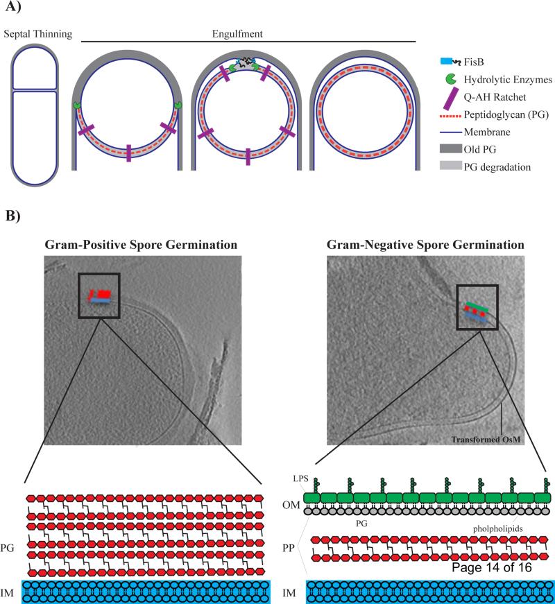
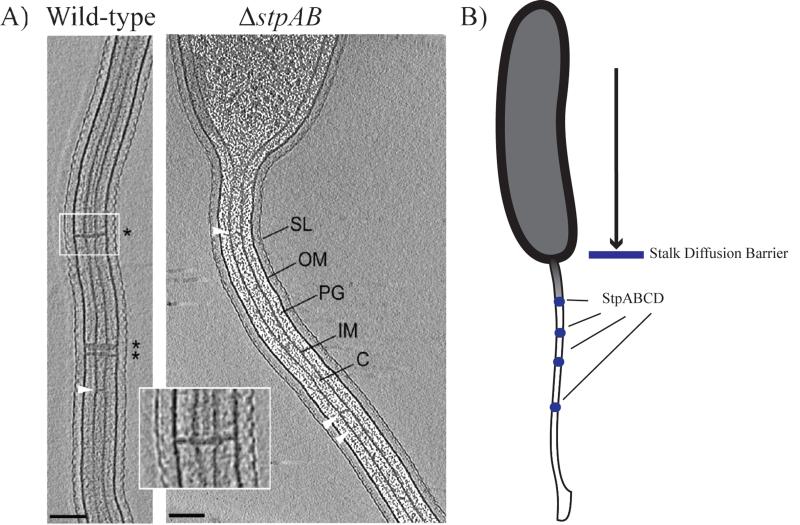
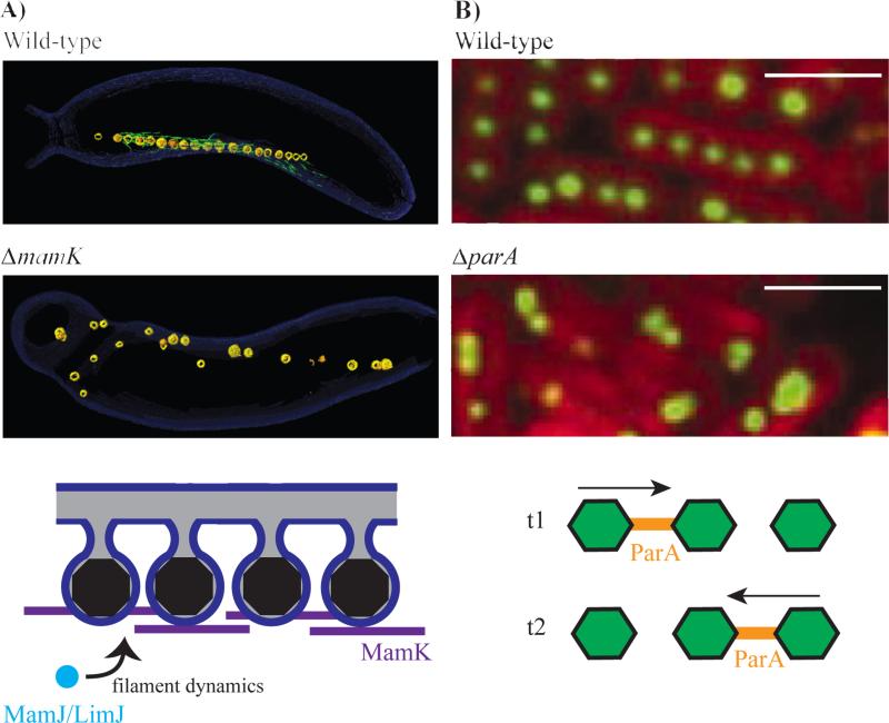
A number of bacterial species rely on compartmentalization to gain specific functionalities that will provide them with a selective advantage. Here, we will highlight several of these modes of bacterial compartmentalization with an eye toward describing the mechanisms of their formation and their evolutionary origins. Spore formation in Bacillus subtilis, outer membrane biogenesis in Gram-negative bacteria and protein diffusion barriers of Caulobacter crescentus will be used to demonstrate the physical, chemical, and compositional remodeling events that lead to compartmentalization. In addition, magnetosomes and carboxysomes will serve as models to examine the interplay between cytoskeletal systems and the subcellular positioning of organelles.


Figures

Figure 1
Figure 1
A) Mechanisms of membrane remodeling during the different stages of engulfment. Engulfment initiates with degradation of septal peptidoglycan, commonly referred to as septal thinning. Peptidoglycan synthesis, peptidoglycan degradation and a specific “ratchet-like” mechanism that is mediated by proteins SpoIIQ and SpoIIIAH (QAH) are all factors that have been shown to be important for driving the mother cell membrane around the forespore. To be released into the mother cell, migrating membranes meet at the cell pole and undergo a fission event that is mediated by FisB. Note that the outer spore membrane (OsM) is derived from the cytoplasmic membrane of the mother cell. B) Spore germination of Gram-positive Bacillus subtilis (left) as compared to Gram-negative Acetonema longum (right). Upon germination, Gram-positive Bacillus subtilis (left) sheds its OsM whereas Gram-negative Acetonema longum (right) retains its OsM. Furthermore, A. longum transforms its OsM into a canonical Gram-negative outer membrane (OM). Cell envelope of Gram-negative vs. Gram-positive bacteria are very different. Gram-positive bacteria have a thick cell wall that is made up of multiple layers of peptidoglycan (PG) that surrounds an inner membrane (IM). On the other hand, Gram-negative bacteria maintain a thin layer of cell wall in-between two membranes. The OM of Gram-negative bacteria is compositionally different from the IM and creates a compartment, the periplasm (PP), that chemically distinct from the cytoplasm. The OM is an asymmetric bilayer with lipopolysaccharide (LPS) distributed in the outer leaflet and phospholipids distributed in the inner leaflet. Copyright (2013) Wiley. Used with permission from [Tocheva et al., Mol Micro, 2013].
Figure 2
Figure 2
A) Electron cryotomography images of wild-type Caulobacter crescentus cells containing stalk diffusion barriers (* and inset). ΔstpAB cells lack these structures. White arrows indicate unidentified structures that span the interior of the stalk (SL= S layer, OM= outer membrane, PG= peptidoglycan, IM= inner membrane, C= cytoplasm). Scale bar 100 nm. Reprinted from [Schlimpert et al., Cell, 2011] with permission from Elsevier. B) Stalk diffusion barriers: Comprising a protein complex of StpA, StpB, StpC and StpD (blue circles), diffusion barriers limit soluble and membrane protein diffusion into the stalk of C. crescentus.
Figure 3
Figure 3
Organization of bacterial organelles by cytoskeletal filaments. A) The bacterial actin protein MamK is responsible for alignment of magnetosomes into a chain in Magentotactic Bacteria. Reconstructed electron cryotomography images of wild-type Magnetospirillum magneticum AMB-1 with an aligned magnetosome chain (top panel) and ΔmamK cells with disorganized magnetosomes (middle panel). The magnetosome (yellow) chain contains iron-based crystals (orange) and is flanked by filamentous structures (green) that disappear in the ΔmamK mutant. MamK dynamic filament behavior is influenced by the presence of MamJ and LimJ (bottome panel). The precise mechanisms that govern this process remain elusive. From [Komeili et al. Sceince, 2006]. Reprinted with permission from AAAS. B) Alignment and segregation of carboxysomes in cyanobacteria is dependent on ParA, another bacterial cytoskeletal protein. Carboxysomes are evenly distributed throughout the cell in wild-type Synechococcus elongatus PCC 7942 (top panel), but not in a mutant lacking parA (middle panel). Rubisco protein (RbcL) fused to YFP indicates the localization of carboxysomes (green) and thylakoid membrane fluorescence is shown in red. ParA oscillates from pole to pole and is found distributed in between carboxysomes (bottom panel). T1 and T2 represent time points that that are roughly 30 minutes apart. From [Savage et al., Science, 2010]. Reprinted with permission from AAAS. Scale bar 2 μm.

References

    1. Spitzer J, Poolman B. How crowded is the prokaryotic cytoplasm? FEBS Lett. 2013;587:2094–2098. - PubMed
    1. Murat D, Byrne M, Komeili A. Cell biology of prokaryotic organelles. Cold Spring Harb. Perspect. Biol. 2010;2:a000422. - PMC - PubMed
    1. Kerfeld C a, Heinhorst S, Cannon GC. Bacterial microcompartments. Annu. Rev. Microbiol. 2010;64:391–408. - PubMed
    1. Hsin J, Chandler DE, Gumbart J, Harrison CB, Sener M, Strumpfer J, Schulten K. Self-assembly of photosynthetic membranes. Chemphyschem. 2010;11:1154–9. - PMC - PubMed
    1. Oostergetel GT, van Amerongen H, Boekema EJ. The chlorosome: a prototype for efficient light harvesting in photosynthesis. Photosynth. Res. 2010;104:245–55. - PMC - PubMed

Publication types

Substances