Review
. 2014 Feb;32(2):149-57.
doi: 10.1038/nbt.2783. Epub 2014 Jan 19.

Beyond model antigens: high-dimensional methods for the analysis of antigen-specific T cells

Affiliations
Review

Beyond model antigens: high-dimensional methods for the analysis of antigen-specific T cells

Evan W Newell et al. Nat Biotechnol. 2014 Feb.

Abstract

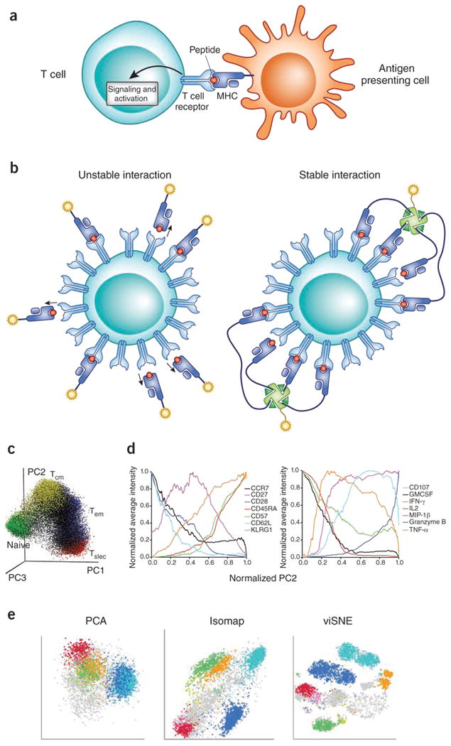
Adaptive immune responses often begin with the formation of a molecular complex between a T-cell receptor (TCR) and a peptide antigen bound to a major histocompatibility complex (MHC) molecule. These complexes are highly variable, however, due to the polymorphism of MHC genes, the random, inexact recombination of TCR gene segments, and the vast array of possible self and pathogen peptide antigens. As a result, it has been very difficult to comprehensively study the TCR repertoire or identify and track more than a few antigen-specific T cells in mice or humans. For mouse studies, this had led to a reliance on model antigens and TCR transgenes. The study of limited human clinical samples, in contrast, requires techniques that can simultaneously survey TCR phenotype and function, and TCR reactivity to many T-cell epitopes. Thanks to recent advances in single-cell and cytometry methodologies, as well as high-throughput sequencing of the TCR repertoire, we now have or will soon have the tools needed to comprehensively analyze T-cell responses in health and disease.


Figures

Figure 1
Figure 1
Antigen recognition by T cell receptor and probing antigen specificity with peptide-MHC multimers. (a) Antigen-specific T cell responses are initiated through the interaction of TCR, expressed on T cells, and the corresponding petide-MHC protein complex expressed by antigen-presenting cells. TCR engagement initiates a complex cell signaling cascade the results in T cell activation. (b) Binding of TCR to its specific peptide-MHC ligand is very low (∼1-100 μM) and has very fast dissociation kinetics (t1/2 usually much less than a minute). Thus, monomeric staining reagents are insufficiently stable for the detection of antigen-specific T cells. In contrast, by taking advantage of cooperative binding, multimeric complexes of peptide-MHC allow for remarkably sensitive and accurate detection of antigen-specific T cells,. (c) Mass cytometry and dimensionality reduction methods allow integrated analysis of T cell phenotype and function. In the example shown here, to visualize diversity of the peripheral blood CD8+ T cells, 25 parameters were measured for each cell including 16 phenotypic markers and 9 functional markers. Principal component analysis (PCA) was applied to generate three aggregate parameters describing ∼60% of total variance. A representative donor's 3D-PCA plot with cells annotated based on previously defined stringent criteria for naïve, central-memory (Tcm), effector-memory (Tem) and short-lived effector cells (Tslec). Adapted from ref . (d) To illustrate the phenotypic and functional meaning of a non-naïve cell progression along the PC2 axis of the PCA plot, average expression of (left) phenotypic and (right) functional parameters were normalized and plotted as a function of normalized PC2 values. This unsupervised analysis provides a hypothetical framework for graded T cell differentiation involving progressive gains and/or losses of surface marker expression and functional capacities. Adapted from ref . (e) To illustrate the power of non-linear dimensionality approaches, a linear PCA analysis of bone-marrow derived cells colored by a number of user-defined cell subsets is compared to two different non-linear approaches, Isomap and viSNE. Adapted from ref .
Figure 2
Figure 2
Single cell analysis can reveal heterogeneity in gene expression among T cells. The data shown here were generated using high-throughput BioMark™ microfluidics for multiplex quantitative RT-PCR analysis. (a) After some careful optimization relative single cell gene expression can be validated by plotting compared to relative expression levels obtained from ‘bulk’ analysis of 100 cells. Good correspondence indicates reliable single cell gene expression measurements. (b) The value of single cell analysis is illustrated by comparing expression of CXCR5 and CCL5 mRNA in single T cells and in ‘bulk’ populations of 100 T cells. Averaging expression of genes across 100 cells masks diversity revealed at the single-cell level. (c) Further illustrating the power of the single-cell approach, expression levels of 24 genes (columns) in 163 cells (rows) is shown. In this example, half of the cells were stimulated prior to analysis (noted as ‘Activated’); the other half were unstimulated (‘Resting’). Unsupervised clustering discriminated all but six cells (noted with X on y-axis) into activated vs. resting categories based on gene expression. Adapted from ref .
Figure 3
Figure 3
Highly mulitplexed analysis of T–cell antigen-specificity using mass cytometry based combinatorial peptide–MHC tetramer staining. (a-b) By coding each T cell antigen specificity (each pMHC tetramer) by a unique combination of three out ten possible metal tags, up to 120 different antigen specificities can be simultaneously measured in a single sample. Cells staining positive with three and only three of the metals comprising each code can be identified as specific for the pMHC epitope corresponding to that code. Adapted from NBT 31:609. (c) This example illustrates the identification of EBV-BRLF-1-specific T cells (colored green) by virtue of their staining positive for 161Dy, 169Tm, and 175Lu after incubation with a pMHC tetramer barcoded with these metals. This panel shows just one of the 120 possible 3D dot-plots for each sample. Adapted from ref . (d) If T cells are also stained with antibodies specific for phenotypic and functional markers, PCA dimensionality reduction can then be used to further summarize the phenotype of T cells specific for each epitope. In this example, each dot represents T cells specific for the indicated epitope from one of the 17 different donors analyzed. T cells displaying previously defined phenotypes and specific for well-characterized antigens can be used as ‘landmarks’ on these plots to help classify the composite phenotype of T cells specific for previously uncharacterized antigens. Adapted from ref . (e) This example illustrates the power of this approach to accurately define the status of antigen-specific T cells. Here the phenotypic status of EBV-specific cells targeting lytic- (BRLF1, BLMF1) versus latency-associated (LMP1, LMP2) antigens are delineated suggesting that T cells specific for lytic-cycle antigens have encountered antigen more recently. Adapted from ref .
Figure 4
Figure 4
Strategies for high-throughput single-cell analysis of TCR sequences and identification of TCR ligands. (a) As demonstrated for B cell immunologlobulin genes, sequencing of V regions of endogenously paired TCRa and TCRb genes can be performed in high-throughput by mRNA capture and emulsion linkage RT-PCR, or by direct cellular emulsion linkage PCR. Cell-specific barcoded tags can also be introduced at the single-cell stage as a general means of increasing the throughput of this type of approach. (b) Libraries of yeast clones displaying 10-10 unique peptides tethered to MHC molecules enables high-throughput screening for peptides capable of binding MHC (because these peptides result in proper folding and surface expression of pMHC complexes) and for peptides capable of binding to a TCR of interest (here the TCR is used as a tetrameric staining reagent). After multiple rounds of selection, hits are sorted, cloned, and sequenced; the peptide sequences can then be analyzed,.

References

    1. Burnet FM. A Modification of Jerne's Theory of Antibody Production using the Concept of Clonal Selection. The Australian Journal of Science. 1957;20:67–69. - PubMed
    1. Burnet FM. The clonal selection theory of acquired immunity. Nashville: Vanderbilt University Press. 1959;3
    1. Hulett HR, Bonner WA, Barrett J, Herzenberg LA. Cell sorting: automated separation of mammalian cells as a function of intracellular fluorescence. Science. 1969;166:747–749. - PubMed
    1. Chattopadhyay PK, Roederer M. Cytometry: today's technology and tomorrow's horizons. Methods. 2012;57:251–258. - PMC - PubMed
    1. Han Q, Bradshaw EM, Nilsson B, Hafler DA, Love JC. Multidimensional analysis of the frequencies and rates of cytokine secretion from single cells by quantitative microengraving. Lab on a chip. 2010;10:1391–1400. - PMC - PubMed

Publication types