Review
. 2014 Jan 16;14(1):1576-97.
doi: 10.3390/s140101576.

Hyperpolarized NMR probes for biological assays

Affiliations
Review

Hyperpolarized NMR probes for biological assays

Sebastian Meier et al. Sensors (Basel). .

Abstract

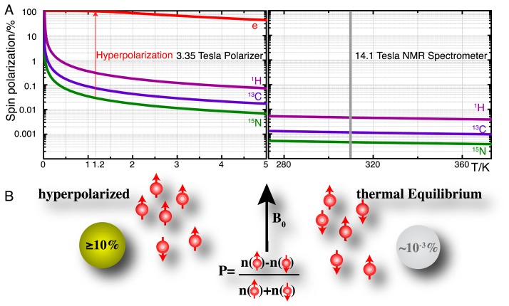
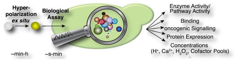
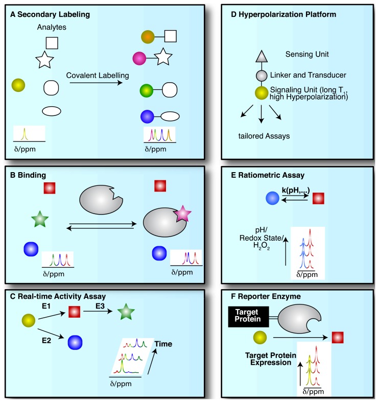
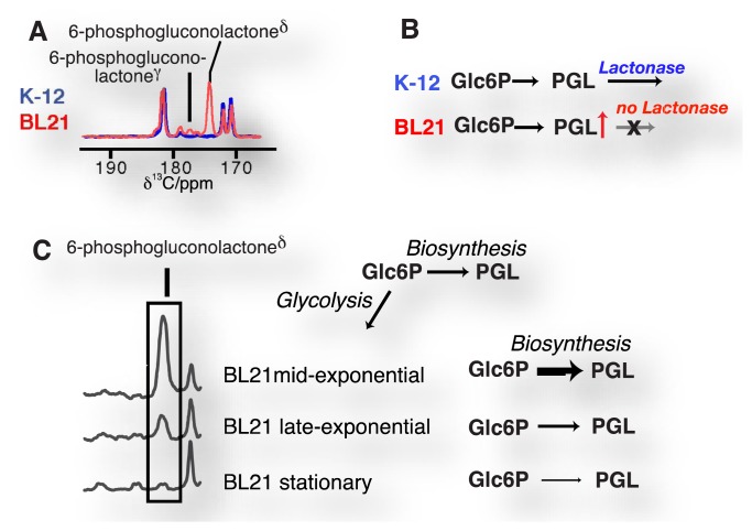
During the last decade, the development of nuclear spin polarization enhanced (hyperpolarized) molecular probes has opened up new opportunities for studying the inner workings of living cells in real time. The hyperpolarized probes are produced ex situ, introduced into biological systems and detected with high sensitivity and contrast against background signals using high resolution NMR spectroscopy. A variety of natural, derivatized and designed hyperpolarized probes has emerged for diverse biological studies including assays of intracellular reaction progression, pathway kinetics, probe uptake and export, pH, redox state, reactive oxygen species, ion concentrations, drug efficacy or oncogenic signaling. These probes are readily used directly under natural conditions in biofluids and are often directly developed and optimized for cellular assays, thus leaving little doubt about their specificity and utility under biologically relevant conditions. Hyperpolarized molecular probes for biological NMR spectroscopy enable the unbiased detection of complex processes by virtue of the high spectral resolution, structural specificity and quantifiability of NMR signals. Here, we provide a survey of strategies used for the selection, design and use of hyperpolarized NMR probes in biological assays, and describe current limitations and developments.


Figures

Figure 1.
Figure 1.
(A) Spin polarizations of electrons (“e”), 1H, 13C and 15N nuclei in a 3.35 Tesla DNP polarizer near liquid helium temperature, compared to spin polarizations of 1H, 13C and 15N in a 14.1 Tesla (600 MHz) spectrometer at 273–373 K. An approach to hyperpolarization is the transfer of electron spin polarization to nuclei near 1.2 K prior to dissolution of the hyperpolarized sample in hot aqueous buffer; (B) resultant hyperpolarized samples in aqueous solutions achieve spin polarizations P that are ∼3–4 orders of magnitude enhanced relative to the thermal equilibrium polarization in an NMR spectrometer.
Figure 2.
Figure 2.
Principle of biological assays using hyperpolarized NMR probes. Hyperpolarization is optimized ex situ and the hyperpolarized probe or label is added to a biomolecule, cell extracts or living cells to conduct biological assays for detection inside an NMR spectrometer.
Figure 3.
Figure 3.
Schematics of different strategies for the use of hyperpolarized labels and probes for NMR spectroscopic biological assays: Hyperpolarized molecules have been used for (A) readout by covalent chemical labeling of analytes; (B) probing of non-covalent binding; (C) the tracking of enzymatic transformations; (D) the design of versatile probe platforms; (E) ratiometric measurements of physicochemical states and (F) interrogating protein expression by probing attached reporter enzymes.
Figure 4.
Figure 4.
Exponential decay time constants for hyperpolarized reporter groups in various designed probes, reaching up to several minutes in symmetrically substituted, non-protonated sites. The reported time constants were derived at 9.4 T and 25 °C for 89Y-DOTP [28], at 14.1 T and 37 °C for permethylated amino acids [51] and at 14.1 T and 30 °C for choline- and TMPA-based probes [38].
Figure 5.
Figure 5.
The direct detection of glucose metabolism in Escherichia coli strains shows the accumulation of a lactone intermediate of the pentose phosphate pathway in strain BL21 (A,B) due to the absence of the lactonase in the BL21 genome, thus affording genomic probing by direct observation of intracellular reaction kinetics; Glc6P = glucose 6-phosphate; PGL = 6-phosphogluconolactone. (C) Accumulation of the lactone occurs in a growth phase dependent manner due to reduced usage of a hyperpolarized glucose probe in biosynthetic pathways as cells approach the stationary phase.
Figure 6.
Figure 6.
Time-resolved observation of metabolite isomers upon feeding a hyperpolarized [2-13C]fructose probe to a Saccharomyces cerevisiae cell cultures at time 0: (A) Glucose 6-phosphate (Glc6P) and fructose 1,6-bisphosphate (Fru1,6P2) C5 signals arise from gluconeogenic reactions of the glycolytic substrate. Isomer ratios are consistent with the formation of the isomers from acyclic intermediates; (B) real-time observation of dihydroxyaceyone phosphate (DHAP) hydrate formation as an off-pathway glycolytic intermediate (other abbreviations are: GA3P = glyceraldehyde 3-phosphate, Ald = aldolase; Pfk = phosphofructokinase; Tpi = triose phosphate isomerase).

References

    1. Palmer A.E., Qin Y., Park J.G., McCombs J.E. Design and application of genetically encoded biosensors. Trends Biotechnol. 2011;29:144–152. - PMC - PubMed
    1. Svatos A. Single-cell metabolomics comes of age: New developments in mass spectrometry profiling and imaging. Anal. Chem. 2011;83:5037–5044. - PubMed
    1. Chan J., Dodani S.C., Chang C.J. Reaction-based small-molecule fluorescent probes for chemoselective bioimaging. Nat. Chem. 2012;4:973–984. - PMC - PubMed
    1. Holman H.Y., Bechtel H.A., Hao Z., Martin M.C. Synchrotron IR spectromicroscopy: Chemistry of living cells. Anal. Chem. 2010;82:8757–8765. - PubMed
    1. Evans C.L., Xie X.S. Coherent anti-stokes raman scattering microscopy: Chemical imaging for biology and medicine. Annu. Rev. Anal. Chem. 2008;1:883–909. - PubMed

LinkOut - more resources