. 2014 Feb;13(2):195-203.
doi: 10.1038/nmat3858. Epub 2014 Jan 19.

Transformation of taxol-stabilized microtubules into inverted tubulin tubules triggered by a tubulin conformation switch

Affiliations

Transformation of taxol-stabilized microtubules into inverted tubulin tubules triggered by a tubulin conformation switch

Miguel A Ojeda-Lopez et al. Nat Mater. 2014 Feb.

Abstract

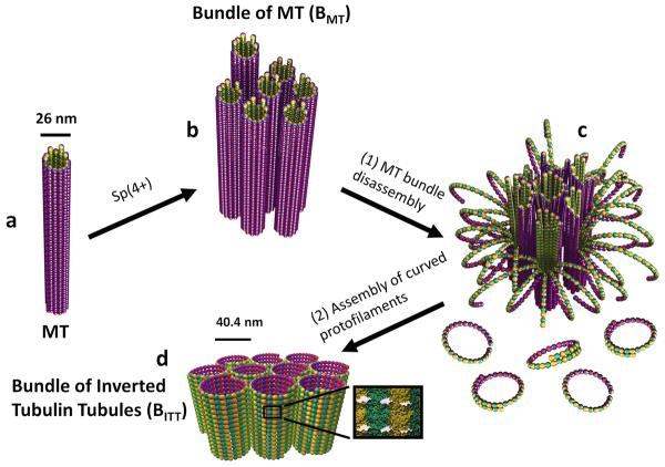
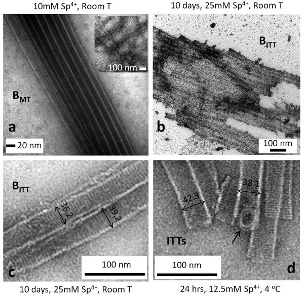
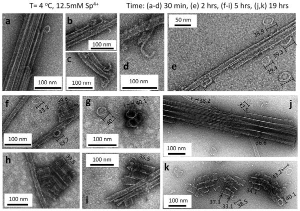
Bundles of taxol-stabilized microtubules (MTs)--hollow tubules comprised of assembled αβ-tubulin heterodimers--spontaneously assemble above a critical concentration of tetravalent spermine and are stable over long times at room temperature. Here we report that at concentrations of spermine several-fold higher the MT bundles (B(MT)) quickly become unstable and undergo a shape transformation to bundles of inverted tubulin tubules (B(ITT)), the outside surface of which corresponds to the inner surface of the B(MT) tubules. Using transmission electron microscopy and synchrotron small-angle X-ray scattering, we quantitatively determined both the nature of the B(MT)-to-B(ITT) transformation pathway, which results from a spermine-triggered conformation switch from straight to curved in the constituent taxol-stabilized tubulin oligomers, and the structure of the B(ITT) phase, which is formed of tubules of helical tubulin oligomers. Inverted tubulin tubules provide a platform for studies requiring exposure and availability of the inside, luminal surface of MTs to MT-targeted drugs and MT-associated proteins.


Figures

Figure 1
Figure 1. Schematic of a spermine (4+)-induced inversion process from bundles of taxol-stabilized microtubules (BMT) to bundles of inverted tubulin tubules (BITT)
a and b, Taxol-stabilized microtubules (MTs, a) may be induced to form MT bundles above a critical concentration of spermine (4+) counterions (BMT, b). The bundles result from the nonspecific electrostatic attraction between spermine coated MTs. c and d, For concentrations several times larger than the critical bundling concentration a specific spermine-triggered straight-to-curved conformation transition in protofilaments, leads to MT disassembly into curved protofilaments (c-PFs) within the bundles (c). Concurrent to MT disassembly spermine counterions induce non-specific assembly of c-PFs into the BITT phase (d). Both phases are hierarchically ordered, liquid crystalline nanotubes, but the tubes are inverted: the tubulin surface, which is on the inside of the tubes in the BMT phase is on the outside in the BITT phase.
Figure 2
Figure 2. TEM of taxol-stabilized microtubule bundles (BMT) and the new spermine-induced phase of bundles of inverted tubulin tubules (BITT)
a, A typical transmission electron microscopy (TEM) image of a taxol-stabilized microtubule bundle (10 mM spermine, room T) showing striations parallel to the cylinder axis due to the protofilaments. The bundle phase is dominant for less than 10 days. Inset, Taxol-stabilized microtubules with straight protofilaments. b and c, An example of a large bundle of inverted tubulin tubules (b, BITT) and a higher magnification of a smaller BITT (c) where protofilaments appear as striations perpendicular to the cylinder axis. TEMs are for 25 mM spermine mixed with taxol-stabilized MTs and imaged after 10 days at room T. d, Example of the BITT phase formed 24 hours after addition of 12.5 mM spermine at 4 °C. Arrow points to overlapping protofilament rings. In this sample preparation a sucrose cushion to remove unpolymerized tubulin (which was used for TEM samples (a–c)) was not employed and inverted tubulin tubules depicted here co-exist with double-walled structures shown in Supplementary Fig. S4 (see Sample Preparation in the Methods section). All TEMs were at taxol/tubulin molar ratio = 0.55.
Figure 3
Figure 3. Time-dependent TEM of the pathway of inversion of taxol-stabilized microtubule bundles (BMT) into bundles of inverted tubulin tubules (BITT) at 4 °C and 12.5 mM spermine
a–d, Transmission electron microscopy (TEM) of early stages of microtubule (MT) bundle disassembly at 30 minutes showing the inside-out curling of protofilaments (PFs) into “pre-ring” structures with a large variation in their diameters (see Supplementary Fig S1). This stage corresponds to Fig. 1c. e, Early-to-intermediate stage TEMs at 2 hrs show the presence of fully formed rings surrounding MT bundles, which dominate the phase at this early-to-intermediate stage where the inverted tubulin structure has not yet formed. TEMs at 1 hr show similar structures. f–i, Intermediate stage TEMs at 5 hrs showing short inverted tubulin tubules (ITTs) and ITT bundles (including an end view of an ITT trimer in g) co-existing with rings, MTs, and MT bundles. j, Late stage TEM at 19 hrs show fully formed bundles of ITTs and few isolated MTs and rings during this late stage. (In the final stage, in the BITT phase, no remaining MTs (and extremely few rings) are found as seen in Fig. 2 both at room T (Fig. 2 b, c) and at 4 °C 24 hrs post addition of 12.5 mM spermine (Fig. 2d).) k, A TEM of a different region of the same sample as in (j) at 19 hrs showing a rare region where short ITTs appear to be forming from the assembly of rings. The variation in the diameter of assembled rings is visible during ITT formation as discussed in the text. The rings in (e–k) have diameters ≈ the diameter of the ITTs. In the measurement of ring size the longer axis was taken because tilts in the ring make it appear as elliptical with the longer axis being a closer estimate of the true diameter. The sample preparations for TEMs (b, c at 30 minutes) and (e–k at 2, 5, 19 hrs) employed a sucrose cushion to remove unpolymerized tubulin after taxol-stabilization of MTs. The sucrose cushion was not employed for TEM samples (a, d at 30 minutes) (see Sample Preparation in the Methods section). All TEMs were at taxol/tubulin molar ratio = 0.55.
Figure 4
Figure 4. Size distribution of ring-like protofilaments in the coexistence regime of disassembling microtubule bundles (BMT) and assembling bundles of inverted tubulin tubules (BITT) from TEM
a–c, Transmission electron microscopy (TEM) images (15 minutes after addition of 15 mM spermine at 0 °C) show coexistence of BMT with short bundles of inverted tubulin tubules (BITT). Also seen are proliferation of ring-like curved protofilaments c-PFs, both in the vicinity of the bundled structures and within disassembling MT bundles (arrows pointing to c-PFs in (b) and (c)). d, Size distribution of 1439 rings in a larger part of the sample surrounding the region shown in the TEM in (a) (see Supplementary Fig. S2). The total number of rings in each box (with a width of 3 nm) is indicated at the top of each box. The mean size of 38.6 nm with a standard deviation of 4.9 nm for the rings is consistent with the diameter of ITTs (≈ 40.4 nm) measured with SAXS of undistorted bundles of ITTs in solution. All TEMs were at taxol/tubulin molar ratio = 0.55.
Figure 5
Figure 5. Synchrotron SAXS data of taxol-stabilized microtubule bundles (BMT) and bundles of inverted tubulin tubules (BITT)
a, Bottom profile shows synchrotron small-angle-x-ray-scattering (SAXS) data of taxol-stabilized microtubules (MT). The second through fourth profiles from bottom are SAXS data from room temperature samples taken 10 days after mixing increasing amounts of spermine with MTs: 5 mM spermine shows 2D hexagonal bundles of MTs (BMT); 15 mM spermine shows coexistence of the BMT phase with the new phase of bundles of inverted tubulin tubules (BITT, arrow points to first order diffraction peak); and 30 mM spermine shows the BITT phase. Top profile shows SAXS of the BITT phase formed at ≈ 2.5 ± 1.5 °C 12 hours after placing microtubules in the BMT phase, with 2.5 mM spermine. b, Three scattering profiles from (a) (bottom two and top curves) after background subtraction with fitted model scattering curves (solid lines) as described in the text. Twelve peaks of the BITT phase can be indexed to a 2D hexagonal lattice. c, Expanded high q region showing comparison of scattering data to a model where the inverted tubulin columns consist of either helical protofilaments with a tight pitch (solid line, which fits the data well) or stacks of rings of curved protofilaments (dotted line, which does not fit the data). All samples were at taxol/tubulin molar ratio = 0.55.
Figure 6
Figure 6. Time-dependent synchrotron SAXS data of the transition kinetics from bundles of microtubules (BMT) to bundles of inverted tubulin tubules (BITT)
a, The BMT phase was suddenly taken from room temperature to ≈ 0° C and the resulting transition to the BITT phase was followed in real time (t) by synchrotron small-angle-xray-scattering (SAXS). The profiles are for SAXS scans of a sample with 15 mM spermine taken at the temperature change (t = 0), thirteen minutes after the temperature change, and subsequently every ten minutes. The scans are offset for clarity with t = 0 at the bottom and t = 93 minutes at the top. b, The amplitude of the (10) peak of the BMT phase (open diamonds) and the BITT phase (open squares) obtained from best fits for the data in (a). For simplicity, the (10) peak of the coexisting BMT and the BITT phases were fit to Gaussians. c, Rates of disassembly of the BMT phase (R(BMT), open diamond) and creation of the BITT phase (R(BITT), open squares) as a function of spermine concentration obtained from fits to SAXS data (as in (a) and (b) for 15 mM spermine). All SAXS samples were at taxol/tubulin molar ratio = 0.55.

Comment in

References

    1. Bray D. Cell Movements: From Molecules to Motility. 2 Garland; New York: 2001.
    1. Thomas TD, Earnshaw WC, Lippincott-Schwartz J. Cell Biology. 2 Saunders, Elsevier; Philadelphia: 2008.
    1. Israelachvili JN. Intermolecular & Surface Forces. Academic Press; Waltham: 1992.
    1. Sackmann E. Membrane Bending Energy Concept of Vesicle-Shape and Cell-Shape and Shape-Transitions. Febs. Letters. 1994;346:3–16. - PubMed
    1. Chiruvolu S, et al. A Phase of Liposomes with Entagled Tubular Vesicles. Science. 1994;266:1222–1225. - PubMed

Publication types