

Visualizing structural dynamics of thylakoid membranes
- PMID: 24442007
- PMCID: PMC3895878
- DOI: 10.1038/srep03768
Visualizing structural dynamics of thylakoid membranes
Abstract
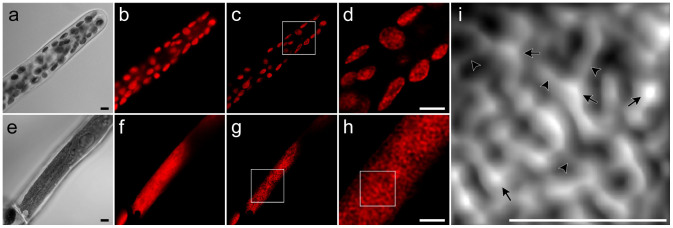
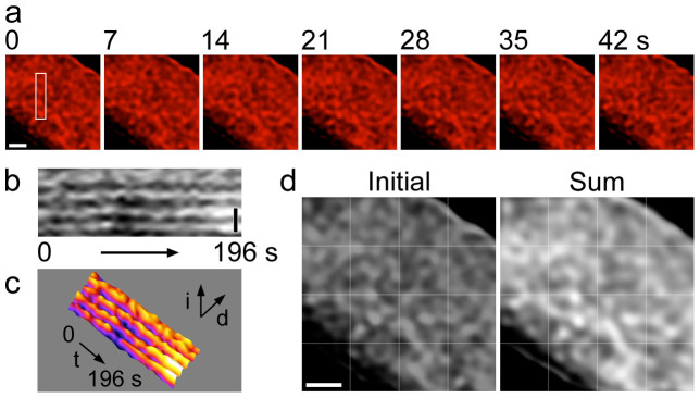
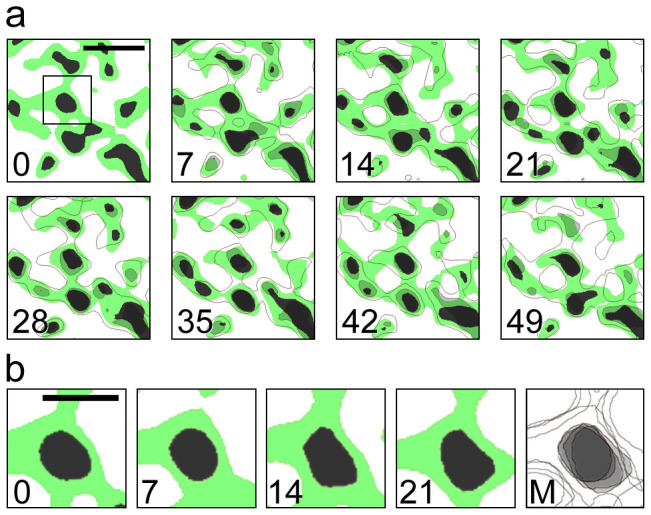
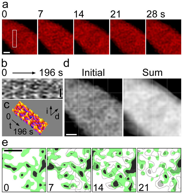
To optimize photosynthesis, light-harvesting antenna proteins regulate light energy dissipation and redistribution in chloroplast thylakoid membranes, which involve dynamic protein reorganization of photosystems I and II. However, direct evidence for such protein reorganization has not been visualized in live cells. Here we demonstrate structural dynamics of thylakoid membranes by live cell imaging in combination with deconvolution. We observed chlorophyll fluorescence in the antibiotics-induced macrochloroplast in the moss Physcomitrella patens. The three-dimensional reconstruction uncovered the fine thylakoid membrane structure in live cells. The time-lapse imaging shows that the entire thylakoid membrane network is structurally stable, but the individual thylakoid membrane structure is flexible in vivo. Our observation indicates that grana serve as a framework to maintain structural integrity of the entire thylakoid membrane network. Both the structural stability and flexibility of thylakoid membranes would be essential for dynamic protein reorganization under fluctuating light environments.
Figures
Similar articles
-
Toward understanding the multiple spatiotemporal dynamics of chlorophyll fluorescence.Plant Signal Behav. 2015;10(6):e1022014. doi: 10.1080/15592324.2015.1022014. Plant Signal Behav. 2015. PMID: 26176900 Free PMC article.
-
Light-Harvesting Complex Stress-Related Proteins Catalyze Excess Energy Dissipation in Both Photosystems of Physcomitrella patens.Plant Cell. 2015 Nov;27(11):3213-27. doi: 10.1105/tpc.15.00443. Epub 2015 Oct 27. Plant Cell. 2015. PMID: 26508763 Free PMC article.
-
Heterologous expression of moss light-harvesting complex stress-related 1 (LHCSR1), the chlorophyll a-xanthophyll pigment-protein complex catalyzing non-photochemical quenching, in Nicotiana sp.J Biol Chem. 2015 Oct 2;290(40):24340-54. doi: 10.1074/jbc.M115.668798. Epub 2015 Aug 10. J Biol Chem. 2015. PMID: 26260788 Free PMC article.
-
Molecular recognition in thylakoid structure and function.Trends Plant Sci. 2001 Jul;6(7):317-26. doi: 10.1016/s1360-1385(01)02010-6. Trends Plant Sci. 2001. PMID: 11435171 Review.
-
Lateral heterogeneity of plant thylakoid protein complexes: early reminiscences.Philos Trans R Soc Lond B Biol Sci. 2012 Dec 19;367(1608):3384-8. doi: 10.1098/rstb.2012.0060. Philos Trans R Soc Lond B Biol Sci. 2012. PMID: 23148264 Free PMC article. Review.
Cited by
-
Toward understanding the multiple spatiotemporal dynamics of chlorophyll fluorescence.Plant Signal Behav. 2015;10(6):e1022014. doi: 10.1080/15592324.2015.1022014. Plant Signal Behav. 2015. PMID: 26176900 Free PMC article.
-
Self-assembly and structural-functional flexibility of oxygenic photosynthetic machineries: personal perspectives.Photosynth Res. 2016 Jan;127(1):131-50. doi: 10.1007/s11120-015-0192-z. Photosynth Res. 2016. PMID: 26494196 Review.
-
State transition is quiet around pyrenoid and LHCII phosphorylation is not essential for thylakoid deformation in Chlamydomonas 137c.Proc Natl Acad Sci U S A. 2022 Sep 13;119(37):e2122032119. doi: 10.1073/pnas.2122032119. Epub 2022 Sep 6. Proc Natl Acad Sci U S A. 2022. PMID: 36067315 Free PMC article.
-
Thylakoid membrane reorganizations revealed by small-angle neutron scattering of Monstera deliciosa leaves associated with non-photochemical quenching.Open Biol. 2020 Sep;10(9):200144. doi: 10.1098/rsob.200144. Epub 2020 Sep 16. Open Biol. 2020. PMID: 32931722 Free PMC article.
-
Protocols for Generating Surfaces and Measuring 3D Organelle Morphology Using Amira.Cells. 2021 Dec 27;11(1):65. doi: 10.3390/cells11010065. Cells. 2021. PMID: 35011629 Free PMC article.
References
-
- Eberhard S., Finazzi G. & Wollman F. A. The dynamics of photosynthesis. Annu. Rev. Genet. 42, 463–515 (2008). - PubMed
-
- Li Z., Wakao S., Fischer B. B. & Niyogi K. K. Sensing and responding to excess light. Annu. Rev. Plant Biol. 60, 239–260 (2009). - PubMed
-
- Allen J. F. & Forsberg J. Molecular recognition in thylakoid structure and function. Trends Plant Sci. 6, 317–326 (2001). - PubMed
Publication types
MeSH terms
Substances
LinkOut - more resources
Full Text Sources
Other Literature Sources
Molecular Biology Databases
