Review
. 2014 Jun;71(12):2313-24.
doi: 10.1007/s00018-014-1557-8. Epub 2014 Jan 18.

Mitochondria dynamism: of shape, transport and cell migration

Affiliations
Review

Mitochondria dynamism: of shape, transport and cell migration

André Ferreira da Silva et al. Cell Mol Life Sci. 2014 Jun.

Abstract

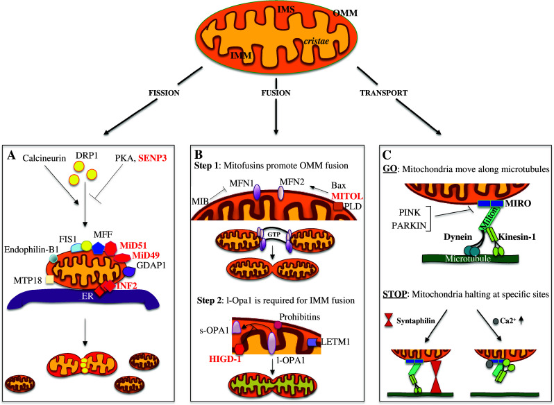
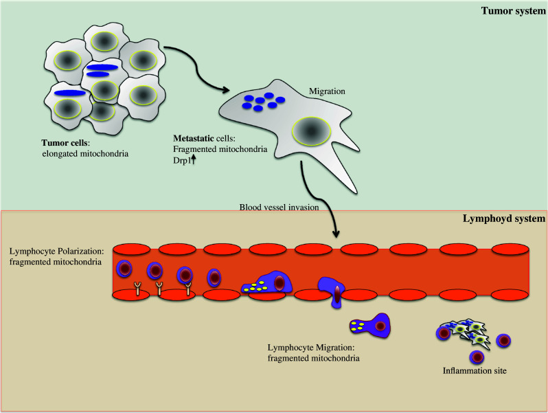
Mitochondria are highly dynamic and functionally versatile organelles that continuously fragment and fuse in response to different physiological needs of the cell. The list of proteins that strictly regulate the morphology of these organelles is constantly growing, adding new players every day and new pieces to the comprehension and elucidation of this complex machinery. The structural complexity of mitochondria is only paralled by their functional versatility. Indeed, changes in mitochondria shape play critical roles in vertebrate development programmed cell death and in various processes of normal cell physiology, such as calcium signaling, reactive oxygen species production, and lifespan. Here, we present the latest findings on the regulation of mitochondrial dynamics and some of their physiological roles, focusing on cell migration. In cells where migration represents a crucial function in their physiology, such as T and tumoral metastatic cells, mitochondria need to be fragmented and recruited to specific subcellular regions to make movement possible. In depth analysis of this role of mitochondrial dynamics should help in identifying potential targeted therapy against cancer or in improving the immune system's efficiency.


Figures

Fig. 1
Fig. 1
Novel players in mitochondrial dynamics regulation. Mitochondria consist of an outer (OMM) and an inner mitochondrial membrane (IMM) that are divided by an inter membrane space (IMS); the IMM can then form narrow tube-like structures called cristae. The balance between the fusion and fission events determines the mitochondria final structure. In the lower panels (ac) the main mitochondria-“shaping-proteins” involved in mitochondria dynamics are represented (labeled in red the most recent). a Model for the fission event. Calcineurin, PKA and SENP3 alternatively control DRP1 localization, promoting (calcineurin) or inhibiting (PKA and SENP3) its translocation to the OMM; MFF, MiD49, and MiD51, all OMM proteins, are independently involved in DRP1 recruitment and regulation of its GTPase activity. Other proteins required for DRP1 recruitment are: FIS1, MTP18, Endophilin-B1, GDAP1, and MFF. The ER and INF2 protein encircles mitochondria marking the future sites of constrictions. DRP1 assemblies at these loci and promotes the fission event. b Mitochondria fusion is a multistep event that initiates with the trans-interaction between the mitofusins proteins, MFN2 and MFN1, positively or negatively regulated by MITOL and MIB, respectively. Once the fusion of the OMM has occurred, OPA1 drives the fusion of the IMM. HIGD-1 is a new player in mitochondria fusion stabilizing the l-OPA1 protein. Additional proteins, such as LETM1, PLD, and Prohibitins, are also involved in the regulation of mitochondrial fusion. c Schematic representation of the proteins required for mitochondrial movement along the microtubules. The mitochondrial adaptor complex, consisting of MIRO and MILTON, acts as a bridge to connect mitochondria with the microtubules. Kinesin-1 and Dynein are the motor proteins regulating the anterograde and retrograde movement, respectively. Different mechanisms are involved in controlling mitochondrial trafficking. Here are presented two processes promoting mitochondria arrest in neurons: SYNTAPHILIN, exclusively axonal, anchors mitochondria through interactions with microtubules; instead, Ca2+, binding to MIRO, determines a rearrangement of the complex so that the motor protein loses its association with the microtubules
Fig. 2
Fig. 2
Mitochondria dynamics modulate cell migration. In the upper part, a schematic representation of a tumor system has been presented. In contrast to tumor cells, metastatic cells have increased levels of DRP1 and fragmented mitochondria. Fragmented mitochondria specifically localized at the uropod of a polarized cell are also hallmarks of T lymphocyte migration (lower part). Thus, in both cell types, the mitochondrial dynamics, and consequently the relocation of mitochondria at specific subcellular compartments, are crucial for the modulation of cell migration/invasion

References

    1. Otera H, Ishihara N, Mihara K. New insights into the function and regulation of mitochondrial fission. Biochim Biophys Acta. 2013;1833:1256–1268. doi: 10.1016/j.bbamcr.2013.02.002. - DOI - PubMed
    1. Tait SW, Green DR. Mitochondria and cell signalling. J Cell Sci. 2012;125:807–815. doi: 10.1242/jcs.099234. - DOI - PMC - PubMed
    1. Baughman JM, Perocchi F, Girgis HS, Plovanich M, Belcher-Timme CA, Sancak Y, Bao XR, Strittmatter L, Goldberger O, Bogorad RL, Koteliansky V, Mootha VK. Integrative genomics identifies MCU as an essential component of the mitochondrial calcium uniporter. Nature. 2011;476:341–345. doi: 10.1038/nature10234. - DOI - PMC - PubMed
    1. De Stefani D, Raffaello A, Teardo E, Szabo I, Rizzuto R. A forty-kilodalton protein of the inner membrane is the mitochondrial calcium uniporter. Nature. 2011;476:336–340. doi: 10.1038/nature10230. - DOI - PMC - PubMed
    1. Pizzo P, Drago I, Filadi R, Pozzan T. Mitochondrial Ca(2)(+) homeostasis: mechanism, role, and tissue specificities. Pflugers Arch. 2012;464:3–17. doi: 10.1007/s00424-012-1122-y. - DOI - PubMed

Publication types

LinkOut - more resources