Review
. 2014 Aug;71(15):2787-814.
doi: 10.1007/s00018-013-1550-7. Epub 2014 Jan 19.

Neuronal calcium signaling: function and dysfunction

Affiliations
Review

Neuronal calcium signaling: function and dysfunction

Marisa Brini et al. Cell Mol Life Sci. 2014 Aug.

Abstract

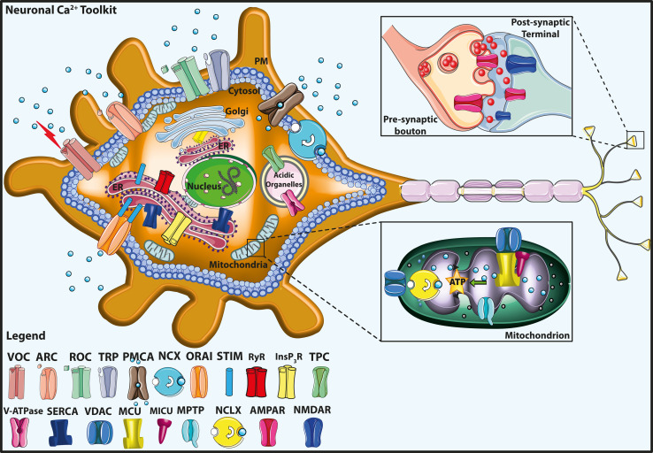
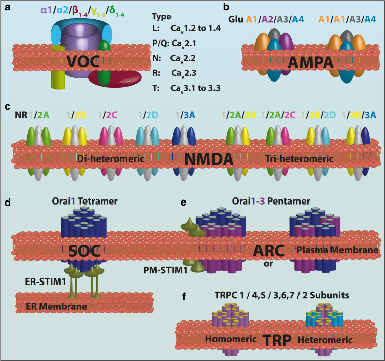
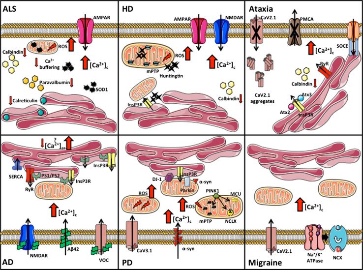
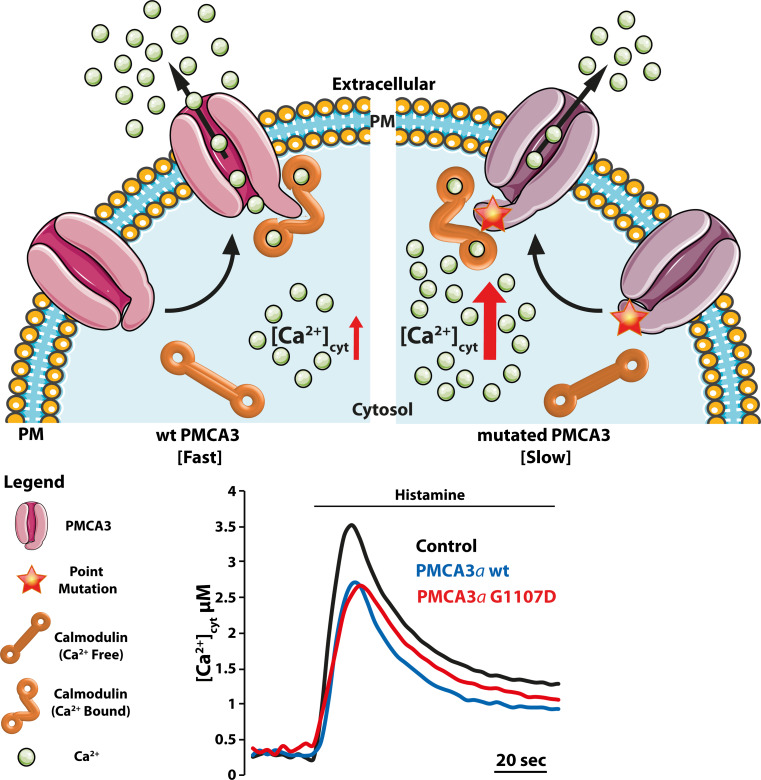
Calcium (Ca(2+)) is an universal second messenger that regulates the most important activities of all eukaryotic cells. It is of critical importance to neurons as it participates in the transmission of the depolarizing signal and contributes to synaptic activity. Neurons have thus developed extensive and intricate Ca(2+) signaling pathways to couple the Ca(2+) signal to their biochemical machinery. Ca(2+) influx into neurons occurs through plasma membrane receptors and voltage-dependent ion channels. The release of Ca(2+) from the intracellular stores, such as the endoplasmic reticulum, by intracellular channels also contributes to the elevation of cytosolic Ca(2+). Inside the cell, Ca(2+) is controlled by the buffering action of cytosolic Ca(2+)-binding proteins and by its uptake and release by mitochondria. The uptake of Ca(2+) in the mitochondrial matrix stimulates the citric acid cycle, thus enhancing ATP production and the removal of Ca(2+) from the cytosol by the ATP-driven pumps in the endoplasmic reticulum and the plasma membrane. A Na(+)/Ca(2+) exchanger in the plasma membrane also participates in the control of neuronal Ca(2+). The impaired ability of neurons to maintain an adequate energy level may impact Ca(2+) signaling: this occurs during aging and in neurodegenerative disease processes. The focus of this review is on neuronal Ca(2+) signaling and its involvement in synaptic signaling processes, neuronal energy metabolism, and neurotransmission. The contribution of altered Ca(2+) signaling in the most important neurological disorders will then be considered.


Figures

Fig. 1
Fig. 1
Neuronal calcium (Ca2+) signaling toolkit. The Ca2+ transport proteins, the receptors of the plasma membrane (PM), and the intracellular organelles, including mitochondria, endoplasmic reticulum (ER), Golgi apparatus, and acidic organelles, are indicated. The mitochondrial Ca2+ handling systems (proteins) are shown in greater detail in the bottom right inset. The top right inset shows a schematic view of the pre-synaptic bouton and the post-synaptic termination. The legend on the bottom left indicates the Ca2+ transporter proteins. VOC Voltage-gated Ca2+ channel, ROC receptor-operated Ca2+ channel, ORAI the pore-forming subunit of store-operated Ca2+ entry channel (SOC), STIM the Ca2+ sensor, TPC two-pore channel, ARC arachidonic acid-regulated Ca2+ channel, TRP transient receptor potential channel, PMCA plasma membrane Ca2+ ATPase, V-ATPase vacuolar H+ ATPase, InsP3R inositol 1,4,5 tris–phosphate receptors, RyR ryanodine receptor, NCX plasma membrane Na+/Ca2+ exchanger, SERCA sarco-/endoplasmic reticulum Ca2+ ATPase, MCU mitochondrial Ca2+ uniporter, MICU mitochondrial Ca2+ uniporter regulator, NCLX mitochondrial Na+/Ca2+ exchanger, VDAC voltage-dependent anion channels, MPTP mitochondrial permeability transition pore, AMPAR 2-amino-3-hydroxy-5-methyl-4-isoxazolepropionic acid receptor, NMDAR N-methyl-d-aspartate receptor
Fig. 2
Fig. 2
Assembly and subtypes of the subunits of the main neuronal Ca2+ channels. a Graphic representation of the VOC complex consisting of the main pore forming α1-subunit plus ancillary β-, γ-, and α2-δ-subunits. Different neuronal α1-subunits correspond to the different types of Ca2+ channels identified in native neurons. b The AMPAR is a ionotropic transmembrane receptor for glutamate assembled from a pool of four subunits (GluA1–A4) which share a high degree of sequence identity. The number of subunits in the functional channels is unclear. The permeability to Ca2+ and other cations, such as sodium (Na+) and potassium (K+), is governed by the GluA2 subunit. An AMPAR lacking a GluA2 subunit will be permeable to Na+, K+, and Ca2+, while the presence of the GluA2 subunit will render the channel impermeable to Ca2+. c The NMDAR is a ionotropic glutamate receptor that allows the flow of Na+, and of smaller amounts of Ca2+, into the cell. Seven NMDAR subunits have been identified: GluNR1, GluNR2A–D, GluNR3A, and GluN3B (not shown). The various populations of di-heteromeric and tri-heteromeric NMDARs that are thought to exist in the central nervous system are shown. d, e The basic structure of ORAI1 and ORAI3 proteins. Each has four transmembrane-spanning domains: the different stoichiometry between SOC and ARCs is shown. The SOC pore is formed by a tetramer of ORAI1 subunits, while a pentameric assembly of three ORAI1 subunits and two ORAI3 subunits arranged in two possible conformations forms the ARC channel. Both SOC and ARC are regulated by STIM1; however, SOC activation is regulated by STIM1 in the endoplasmic reticulum (ER), while the pool of STIM1 residing in the plasma membrane regulates the ARC. f Transient receptor potential (TRP) channels (TRPC) are relatively non-selectively permeable to cations, including Na+, Ca2+, and Mg2+. They belong to a large superfamily of cation channels having six transmembrane-spanning segments, which presumably assemble in a ring-like structure with fourfold symmetry. The TRP protein assembles into homo-tetramers or hetero-tetramers. Here, only the “common” family of these TRPC, with its relative subunits, is shown for simplicity
Fig. 3
Fig. 3
Perturbed Ca2+ homeostasis in neuronal disorders. The cartoon summarizes the main pathways involved in the Ca2+ dyshomeostasis processes linked to the neuronal pathologies described in the text. ALS Amyotrophic lateral sclerosis, HD Huntington’s disease, AD Alzheimer’s disease, PD Parkinson’s disease, ROS reactive oxygen species, PS presenilin, SOD superoxide dismutase, mPTP mitochondrial permeability transition pore, ATX ataxin, PINK1 PTEN-induced kinase, α-syn alpha-synuclein
Fig. 4
Fig. 4
Three Parkinson’s disease (PD)-related proteins, namely, α-synuclein (α-syn), parkin, and DJ-1, control mitochondrial Ca2+ uptake by modulating ER–mitochondria contact sites. Mitochondria accumulate Ca2+ into the matrix via the electrophoretic MCU using the electrochemical gradient generated by the respiratory chain. The VDAC controls Ca2+ diffusion through the outer mitochondrial membrane, facilitating Ca2+ entry into mitochondria by coupling with the InsP3R through the chaperone protein Grp75. The NCLX mediates Ca2+ extrusion from the mitochondria, thus preventing the attainment of the thermodynamic equilibrium. ER–mitochondria tethering has recently emerged as a key element in cell Ca2+ metabolism and mitochondrial physiology. Our recent findings provide evidence for a role of α-syn, parkin and DJ-1 in favoring ER–mitochondria Ca2+ transfer by enhancing the ER–mitochondria contact sites [–215]. The traces (right) refer to mitochondrial Ca2+ transients generated by the stimulation of HeLa cells overexpressing parkin, α-syn, or DJ-1 with an InsP3-linked agonist (histamine). The peak amplitude of the mitochondrial Ca2+ transients was higher in overexpressing cells than in control cells
Fig. 5
Fig. 5
A mutation in the plasma membrane (PM) Ca2+ ATPase 3 (PMCA3) pump causes X-linked congenital cerebellar ataxia. The PMCA3 pump is highly expressed in the cerebellum, particularly in the presynaptic terminals of parallel fibers–Purkinje neurons. The cartoon in the figure summarizes the recent finding that a missense mutation of the X-linked PMCA3 pump gene in a family with congenital cerebellar ataxia impaired the Ca2+ extrusion ability of the pump. The figure also shows cytosolic Ca2+ measurements in model cells (HeLa) overexpressing the PMCA3a wild-type (wt) (blue trace) or PMCA3a G1107D mutant pump (red trace). Cells were stimulated with a Ca2+ mobilizing agonist (histamine). As mentioned in the text, the “a” isoform is a spliced variant of the PMCA3 pump. The black trace in the bottom panel shows the Ca2+ transient in untransfected control cells. The reduction of the peak amplitude in PMCA3-overexpressing cells is due to the Ca2+ extrusion activity of the overexpressed pump. The mutant PMCA3a pump (red trace) has a reduced ability to extrude Ca2+ from the cells: the decline of the red trace is slower than that of the blue trace, which corresponds to cells overexpressing the wt PMCA3a pump [69]

References

    1. Carafoli E, Malmstrom K, Sigel E, Crompton M. The regulation of intracellular calcium. Clin Endocrinol (Oxf) 1976;5(Suppl):49S–59S. - PubMed
    1. Brini M, Cali T, Ottolini D, Carafoli E. Intracellular calcium homeostasis and signaling. Met Ions Life Sci. 2013;12:119–168. - PubMed
    1. Mellstrom B, Savignac M, Gomez-Villafuertes R, Naranjo JR. Ca2+-operated transcriptional networks: molecular mechanisms and in vivo models. Physiol Rev. 2008;88(2):421–449. - PubMed
    1. Carafoli E. The unusual history and unique properties of the calcium signal. In: Krebs J, Michalak M, editors. Calcium: a matter of life and death. Amsterdam: Elsevier; 2007. pp. 3–22.
    1. Prakriya M, Feske S, Gwack Y, Srikanth S, Rao A, Hogan PG. Orai1 is an essential pore subunit of the CRAC channel. Nature. 2006;443(7108):230–233. - PubMed

Publication types