. 2014 Feb;3(2):265-75.
doi: 10.5966/sctm.2013-0025. Epub 2014 Jan 17.

Three-year outcomes of cultured limbal epithelial allografts in aniridia and Stevens-Johnson syndrome evaluated using the Clinical Outcome Assessment in Surgical Trials assessment tool

Affiliations

Three-year outcomes of cultured limbal epithelial allografts in aniridia and Stevens-Johnson syndrome evaluated using the Clinical Outcome Assessment in Surgical Trials assessment tool

Alex J Shortt et al. Stem Cells Transl Med. 2014 Feb.

Abstract

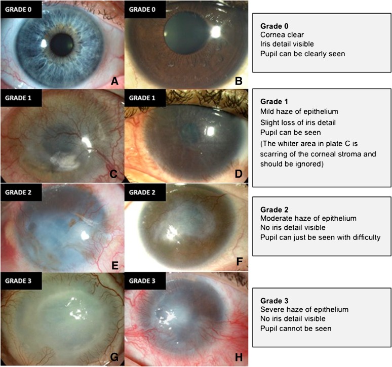
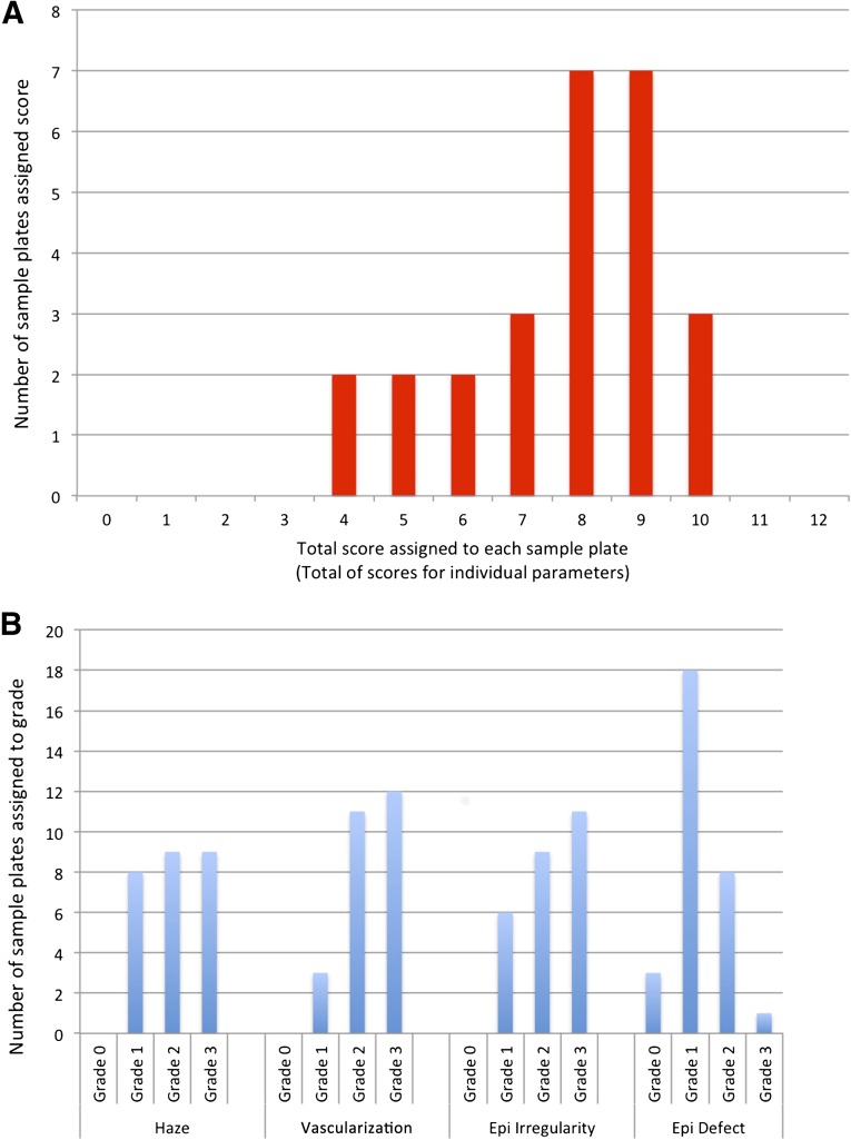
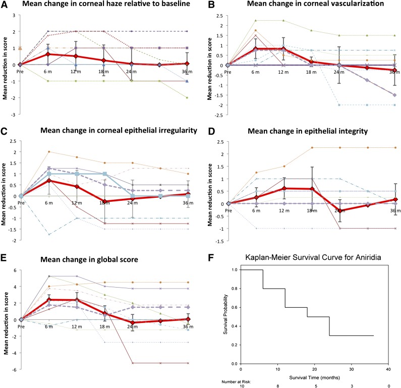
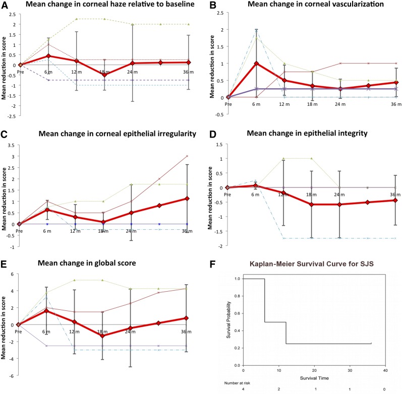
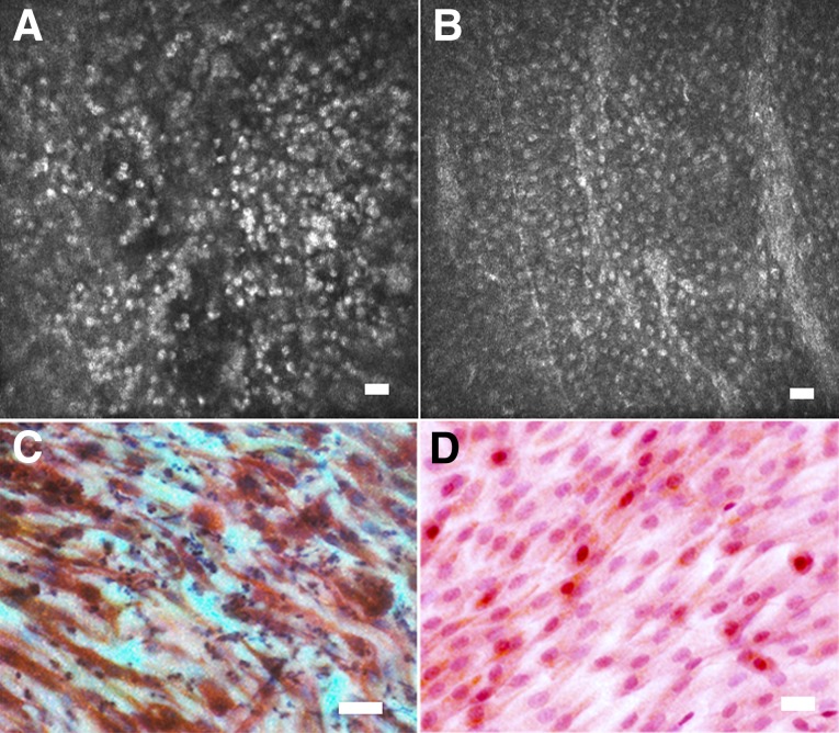
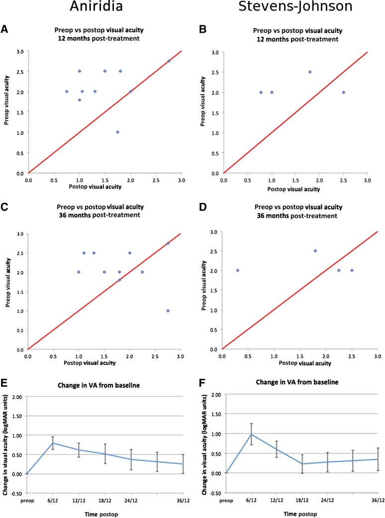
Limbal stem cell deficiency (LSCD) is an eye disorder in which the stem cells responsible for forming the surface skin of the cornea are destroyed by disease. This results in pain, loss of vision, and a cosmetically unpleasant appearance. Many new treatments, including stem cell therapies, are emerging for the treatment of this condition, but assessment of these new technologies is severely hampered by the lack of biomarkers for this disease or validated tools for assessing its severity. The aims of this study were to design and test the reliability of a tool for grading LSCD, to define a set of core outcome measures for use in evaluating treatments for this condition, and to demonstrate their utility. This was achieved by using our defined outcome set (which included the Clinical Outcome Assessment in Surgical Trials of Limbal stem cell deficiency [COASTL] tool) to evaluate the 3-year outcomes for allogeneic ex vivo cultivated limbal epithelial transplantation (allo-CLET) in patients who had bilateral total LSCD secondary to aniridia or Stevens-Johnson syndrome. The results demonstrate that our new grading tool for LSCD, the COASTL tool, is reliable and repeatable, and that improvements in the biomarkers used in this tool correlate positively with improvements in visual acuity. The COASTL tool showed that following allo-CLET there was a decrease in LSCD severity and an increase in visual acuity up to 12 months post-treatment, but thereafter LSCD severity and visual acuity progressively deteriorated.

Keywords: Cornea; Outcome measures; Stem cell; Stem cell deficiency; Stem cell transplantation.


Figures

Figure 1.
Figure 1.
Standardized grading plate used to grade corneal epithelial haze as normal (grade 0, no signs) (A, B), mild (grade 1) (C, D), moderate (grade 2) (E, F), or severe (grade 3) (G, H).
Figure 2.
Figure 2.
Graphs used to assess distribution of grading scores. (A): Distribution of global scores (the sum of the scores assigned to each of the four key signs of limbal stem cell deficiency [LSCD]) for 26 patients with limbal stem cell deficiency. Scores were distributed over a wide range on the scale, reflecting the range of LSCD severities in the population sample. (B): Distribution of scores within individual clinical parameters. Scores again show an even distribution with the exception of grade 1 epithelial defects. Abbreviation: Epi, epithelial.
Figure 3.
Figure 3.
Outcomes of ex vivo cultivated limbal allografts for aniridia. (A–D): Effect of treatment on scores for corneal haze (A), vascularization (B), corneal epithelial irregularity (C), and integrity (epithelial defects) (D). In these graphs the solid red line represents the mean for all patients, the error bars indicate the 95% confidence intervals for the mean, and the dashed lines represent the results for individual patients. (E, F): Kaplan-Meier survival curve (F), where success or survival was defined as a 25% improvement in the global score (E) versus the preoperative global score.
Figure 4.
Figure 4.
Outcomes of ex vivo cultivated limbal allografts for Stevens-Johnson syndrome. (A–D): Effect of treatment on scores for corneal haze (A), vascularization (B), corneal epithelial irregularity (C), and integrity (epithelial defects) (D). In these graphs, the solid red line represents the mean for all patients, the error bars indicate the 95% confidence intervals for the mean, and the dashed lines represent the results for individual patients. (E, F): Kaplan-Meier survival curve (F), where success or survival was defined as a 25% improvement in the global score versus the preoperative global score (E). Abbreviation: SJS, Stevens-Johnson syndrome.
Figure 5.
Figure 5.
Nonstratifying epithelial phenotype observed in in vivo confocal microscopy and impression cytology observed in several patients with limbal stem cell deficiency. Shown are confocal microscopy (A, B) and corresponding impression cytology (C, D) images from the central cornea of two patients with limbal stem cell deficiency prior to treatment. (A, B): Confocal microscopy shows a thin sheet of small cells, usually a mono- or bilayer, with hyperfluorescent nuclei, but no other cellular detail is visible. (C, D): Correlation with impression cytology performed on the same patients directly after confocal scans showed that these cells had a spindle-like shape and expressed CK19 (red) but not CK3 (brown) (C, D). This phenotype may represent conjunctival cell migration onto the corneal stroma but failure of the cell layer to form cell junctions and differentiate into a mature multilayered epithelium. Scale bars = 25 μm.
Figure 6.
Figure 6.
Visual acuity data for patients with aniridia and Stevens-Johnson syndrome treated with ex vivo cultivated limbal allografts. Values are means, and error bars indicate the 95% confidence intervals. Abbreviations: Postop, postoperative; Preop, preoperative; VA, logMAR visual acuity.

References

    1. Daniels JT, Dart JK, Tuft SJ, et al. Corneal stem cells in review. Wound Repair Regen. 2001;9:483–494. - PubMed
    1. Grueterich M, Espana EM, Tseng SC. Ex vivo expansion of limbal epithelial stem cells: amniotic membrane serving as a stem cell niche. Surv Ophthalmol. 2003;48:631–646. - PubMed
    1. Holland EJ. Epithelial transplantation for the management of severe ocular surface disease. Trans Am Ophthalmol Soc. 1996;94:677–743. - PMC - PubMed
    1. Kim JY, Djalilian AR, Schwartz GS, et al. Ocular surface reconstruction: Limbal stem cell transplantation. Ophthalmol Clin North Am. 2003;16:67–77. - PubMed
    1. Kinoshita S, Adachi W, Sotozono C, et al. Characteristics of the human ocular surface epithelium. Prog Retin Eye Res. 2001;20:639–673. - PubMed

Publication types

MeSH terms